13 Facial Danger Zone 4 – Nasolabial Region


10.1055/b-0040-177255

13 Facial Danger Zone 4 – Nasolabial Region

Rod J. Rohrich and Raja Mohan


Abstract


This chapter summarizes how to inject soft-tissue fillers into the nasolabial region. Patients often report a prominent nasolabial fold as they age, and one option for treatment is the injection of soft-tissue filler. The facial artery’s anatomical location is intimately related to the location of the nasolabial fold. We present safe techniques for injection soft-tissue filler into this region to prevent inadvertent injury to any major facial vessels.


Key Points for Maximizing Filler Safety in the Nasolabial Region


  • Use only FDA-approved reversible hyaluronic acid fillers in most areas of the face.


  • Hyaluronic acid fillers are reversible if there is a vascular issue or problem because they can be reversed with hyaluronidase.


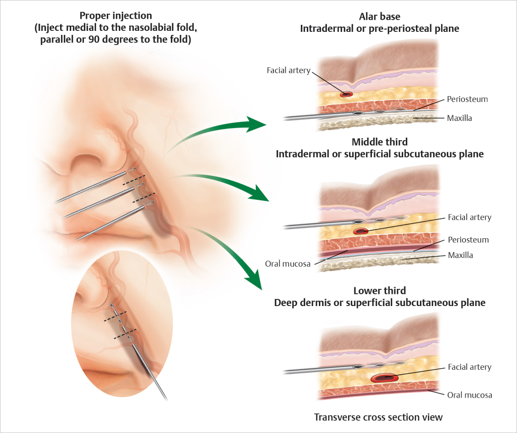
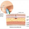
  • In the lower two-thirds of the nasolabial fold, inject into the deep dermis or superficial subcutaneous plane just medial to the nasolabial fold (▶ Fig. 13.1 ).


  • Near the alar base, inject either intradermally or in the preperiosteal plane. Use incremental deep depot injection techniques in the periapical areas (▶ Fig. 13.1 ).


  • Always perform using gentle, low-pressure anterograde/retrograde injections with constant motion in 1 mL syringes.


  • Do not inject along the alar rim, alar grooves, or nasal sidewall because the vasculature is superficial in these regions.

Fig. 13.1 Proper injection technique into nasolabial fold. The key in augmenting the nasolabial fold is to stay medial to the fold to prevent inadvertent injury or injections into the adjacent vasculature. In the upper third of the nasolabial zone, injections should be performed deep in a preperiosteal plane or very superficially in an intradermal plane. The artery is located within the subcutaneous tissue. In the middle third, the artery is located deeper, so injections should be performed intradermally or in a superficial subcutaneous plane. Lastly, in the lower third of the nasolabial zone, the artery is either within the muscle or between the muscle and the subcutaneous tissue, so more superficial injections are recommended.


13.1 Safety Considerations in the Nasolabial Region


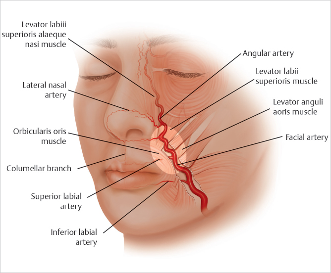
  • When injecting the nasolabial region, knowledge of the depth and course of the facial artery is paramount to prevent complications associated with intravascular injury ( Fig. 13.2).


  • In the lower two-thirds of the nasolabial fold, the facial artery course lies beneath the muscle or in the deeper planes above the muscle (▶ Fig. 13.3 ).


  • The artery becomes superficial at the upper third of the nasolabial fold and is most at risk for injury at this level (▶ Fig. 13.3) (Video 13.1).


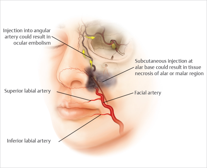
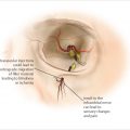
  • Subcutaneous injections in the upper third of the nasolabial fold can lead to soft-tissue necrosis of the alar or malar regions if there is intravascular injury (▶ Fig. 13.4 ).


  • In the upper third of the nasolabial fold and superior to it, intravascular injections into the angular artery could result in ocular embolism (▶ Fig. 13.4 ).


  • The nasolabial fold is the second most common injection site for tissue necrosis and the third most common site leading to visual loss. 1 , 2

Fig. 13.2 Nasolabial danger zone. The nasolabial danger zone is highlighted in the diagram. The tortuous course of the facial artery is also shown. Inferiorly, the artery is located deeper and becomes more superficial nearby the alar base. Its location is closely related to the nasolabial fold, so care must be taken when augmenting the fold. The facial artery has many important branches such as the inferior labial artery, superior labial artery, and lateral nasal artery.
Fig. 13.3 Facial cadaver dissection highlighting details of facial artery anatomy. With the subcutaneous tissue (e) reflected, the facial artery (a) is seen running in the nasolabial fold, at times within the muscle but mostly in the plane between the subcutaneous tissue and muscle. The artery becomes superficial (b) in the upper third of the nasolabial fold and is at risk during superficial injections. The transition of the facial artery into the angular artery (c) and its anastomosis with the dorsal nasal artery (d) is demonstrated. Of note, the facial artery lies approximately 1.5 cm lateral to the commissure.
Fig. 13.4 Risk of nasolabial fold injection. Potential routes for retrograde embolization to the ophthalmic vessels are shown in the figure. Injections superficially near the alar base could result in deposits in the angular artery that can migrate in a retrograde fashion. Superficial injections near the alar base can also result in vascular compromise of the alar and malar soft tissues.

Only gold members can continue reading. Log In or Register to continue

Stay updated, free articles. Join our Telegram channel

Jul 18, 2020 | Posted by in Dermatology | Comments Off on 13 Facial Danger Zone 4 – Nasolabial Region

Full access? Get Clinical Tree

Get Clinical Tree app for offline access