11 Facial Danger Zone 2 – Temporal Region
Abstract
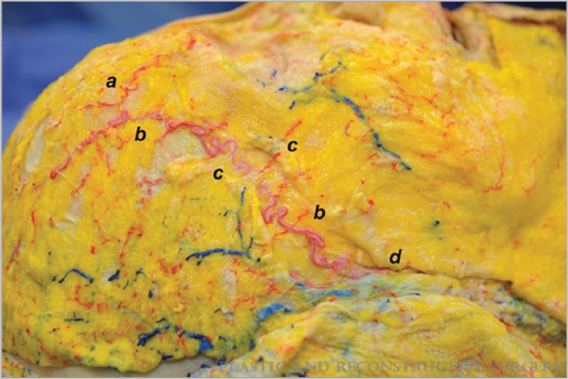
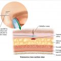
The superficial temporal artery and middle temporal vein lie within the temporal fossa in an intermediate plane. Inadvertent injection into the frontal branch of the superficial temporal artery can cause ocular compromise via retrograde embolization through the supraorbital system. Injection into the middle temporal vein may result in nonthrombotic pulmonary embolism via anterograde venous flow into the internal jugular vein. Filler injections in the temporal region should be performed either superficially in the subcutaneous tissue, or deeply in the preperiosteal plane to avoid inadvertent cannulation of at-risk vessels situated in the intermediate plane.
Key Points for Maximizing Filler Safety in the Temporal Region
Avoid injecting in the intermediate plane where vulnerable vessels lie in the temporal region.
Inject either superficially in the superficial subcutaneous tissue or deeply in the preperiosteal plane.
Inject with low pressure in an anterograde/retrograde motion.
11.1 Safety Considerations in the Temporal Region
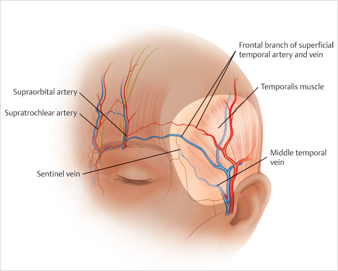
The superficial temporal artery and middle temporal vein lie within the temporal fossa in an intermediate plane (▶ Fig. 11.1 ).
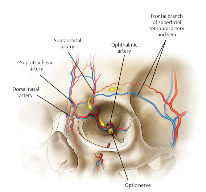
Intravascular injection of foreign material into the frontal branch of the superficial temporal artery can cause ocular compromise via retrograde embolization through the supraorbital system 1 (▶ Fig. 11.2).
Dye injected into the superficial temporal artery has been found within ipsilateral and even bilateral globes in cadaver studies. 2
Although extremely rare, intravascular injection into the middle temporal vein can cause nonthrombotic pulmonary embolism via anterograde venous flow into the internal jugular vein. 3 , 4
11.2 Pertinent Anatomy of the Temporal Region
11.2.1 Superficial Temporal Artery – Frontal Branch
(▶Fig. 11.3) (Video 11.1)
Path is similar to the temporal branch of the facial nerve.
Originates 1 fingerbreadth anterior and 2 fingerbreadths superior to the tip of the tragus. 5
Runs in the intermediate plane within temporoparietal fascia 2 cm above zygomatic arch. 1 , 6
Transitions to the subcutaneous plane 1 fingerbreadth superior to the peak of the brow near the lateral border of the frontalis. 1
Anastomoses with the supraorbital artery above the lateral brow.
Related posts:


Stay updated, free articles. Join our Telegram channel

Full access? Get Clinical Tree


