10 Thumb MCP Joint Ulnar Collateral Ligament Repair
10.1055/b-0038-161078
10 Thumb MCP Joint Ulnar Collateral Ligament Repair
Dariush Nikkhah, Amir H. Sadr
10.1 Ulnar Collateral Ligament Repair
Acute rupture of the metacarpophalangeal joint (MCPJ) ulnar collateral ligament (UCL) also known as Skier thumb is a result of forced abduction that can further result in a UCL tear. The primary role of the MCPJ of the thumb is flexion and extension. The joint is stabilized by the UCL and radial collateral ligament (RCL), the volar plate, and the dorsal capsule. The collateral ligaments consist of a strong proper collateral ligament and a weaker accessory collateral ligament.
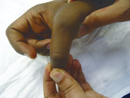
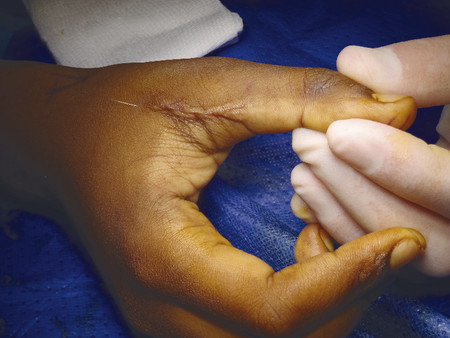
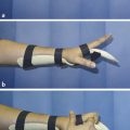
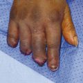
To test for joint stability, one should test by fully flexing the MCPJ and should apply valgus stress to the thumb to test the proper collateral ligament. Laxity of over 35 degrees with no clear endpoint signifies rupture (Fig. 10‑1). Full extension of MCPJ with a valgus stress test assesses the accessory collateral ligament. One should make a comparison with the uninjured contralateral thumb.
Fig. 10.1 Testing proper UCL in MCPJ flexion, no clear endpoint suggestive of UCL tear.
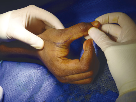
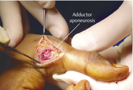
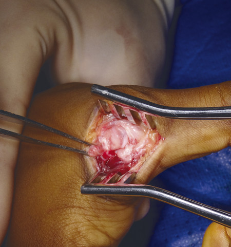
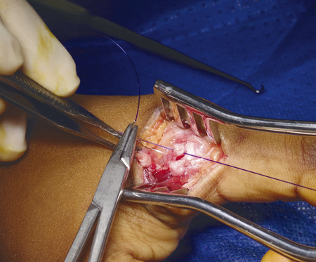
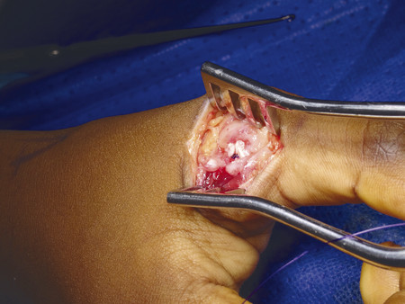
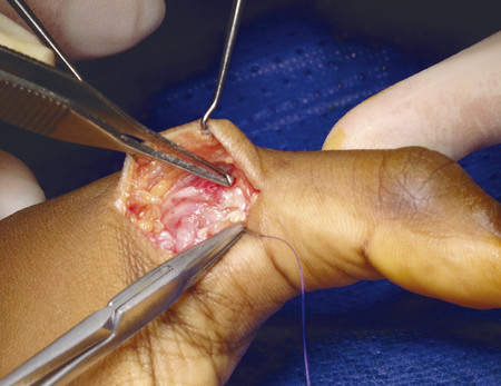
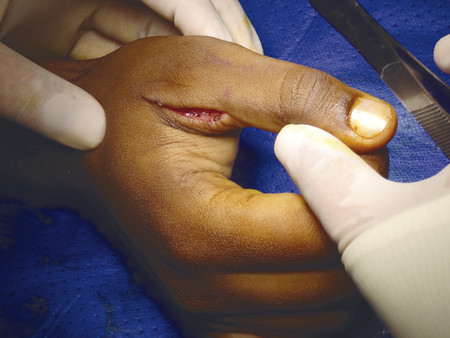
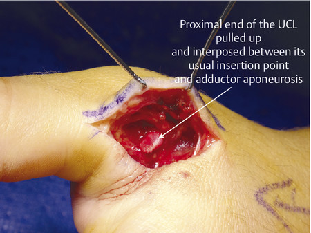
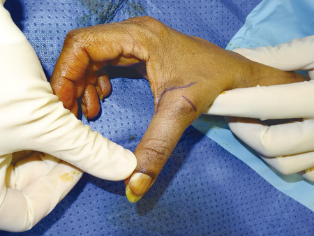
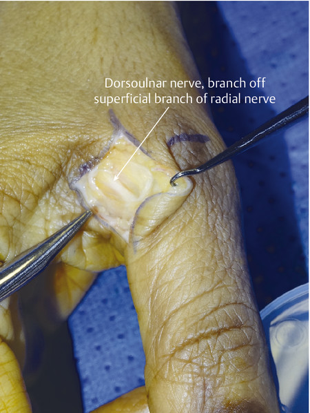
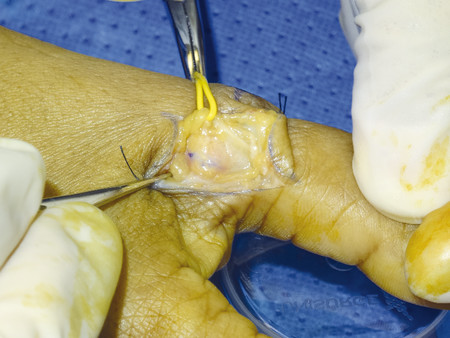
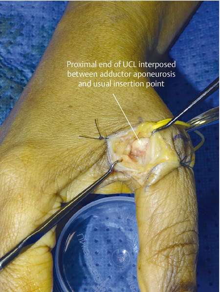
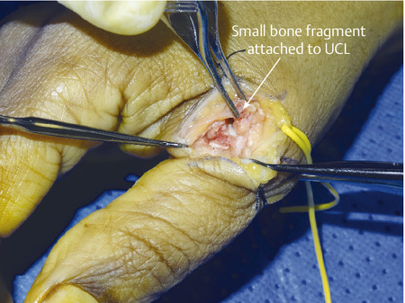
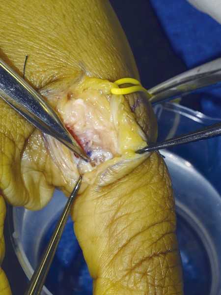
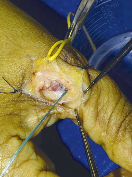
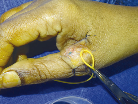
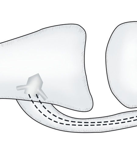
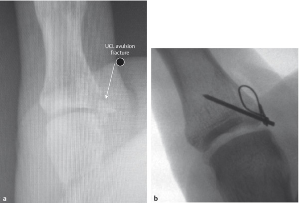
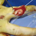
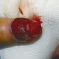
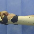
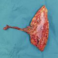
Fig. 10.2 A curvilinear incision is made over the UCL, making sure the incision is marked sufficiently volar to access the UCL.Fig. 10.3 After the skin incision, the adductor aponeurosis and hematoma are visualized; it is important not to divide the terminal branch of the superficial branch of radial nerve (SBRN) and retract it to one side.Fig. 10.4 Once the adductor aponeurosis is opened, access to UCL can be achieved. Here in the figure, the proximal end of the UCL is held with forceps.Fig. 10.5 As this is a midsubstance tear of the UCL, repair is achieved with a 3.0 PDS mattress suture. To provide additional strength, the dorsal joint capsule should also be repaired.Fig. 10.6 The UCL is repaired with the PDS knot visible.Fig. 10.7 Repair of the adductor aponeurosis with a 6.0 PDS running stitch.Fig. 10.8 After the final repair, the thumb is showing no laxity on valgus stress test.Fig. 10.9 Final skin closure with subcuticular 5.0 Monocryl.Fig. 10.10 Stener lesion with proximal end of UCL visible over adductor aponeurosis.Fig. 10.11 Marked laxity of MCPJ suggestive of UCL rupture.Fig. 10.12 Terminal branch of SBRN identified and protected.Fig. 10.13 Retraction of SBRN nerve, cut through adductor aponeurosis as marked in purple ink.Fig. 10.14 Proximal end of UCL curled over adductor aponeurosis—so-called Stener lesion.Fig. 10.15 Small bony segment avulsed off proximal phalanx, bony segment is removed before fixation with Mitek mini anchor onto the proximal phalanx.Fig. 10.16 Drill hole made in the proximal phalanx with white 18-gauge needle to accommodate Mitek mini anchor.Fig. 10.17 Mitek mini anchor fitted into bone and 2.0 Ticron used to pass through UCL as a half-modified Kessler suture.Fig. 10.18 Final repair.Fig. 10.19 Illustration of Mitek mini anchor.Fig. 10.20 (a,b) Tension band wire technique for UCL avulsion with large bony fragment.
Only gold members can continue reading. Log In or Register to continue