, Shimin Chang2, Jian Lin3 and Dajiang Song1
(1)
Department of Orthopedic Surgery, Changzheng Hospital Second Military Medical University, Shanghai, China
(2)
Department of Orthopedic Surgery, Yangpu Hospital Tongji University School of Medicine, Shanghai, China
(3)
Department of Microsurgery, Xinhu Hospital Shanghai Jiao Tong University, Shanghai, China
The distally based lateral supramalleolar flap, nourished by the supramalleolar anterior perforating branch of the peroneal artery, was firstly reported by Masquelet in 1988 [1]. The flap was then modified by other researchers to reach more distal region such as the forefoot defect.
27.1 Vascular Anatomy
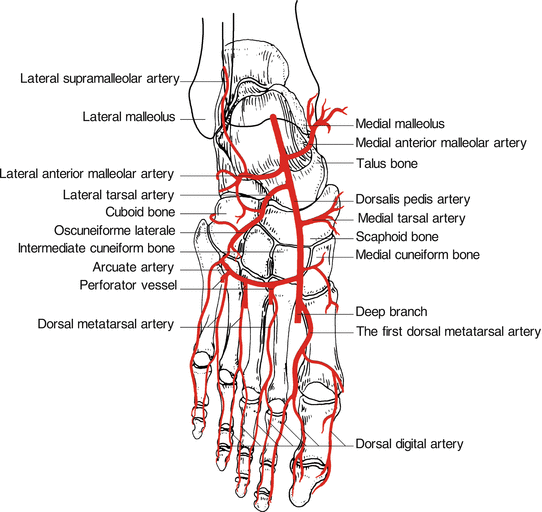
The anterolateral supramalleolar flap is based on the anterior perforating branch of the peroneal artery, which pierces the intermuscular septum and appears on the front of the leg approximately 5 cm above the lateral malleolus. Here it divides into 2 branches, one is a deep branch which runs distally in the loose areolar fatty tissue under the deep fascia (retinaculum), running over the tarsal bones and anastomosing with other vessels, such as the branches of the anterior tibial artery, dorsalis pedis artery, and the terminal part of the peroneal artery which is called lateral calcaneal artery. The other branch is a superficial cutaneous branch, which emerges between the extensor digitorum longus and the peroneus brevis muscles and expended in proximal direction.
The flap pedicle can be designed in 2 versions. One is based on the perforating point with a local transposition for malleolar defect. The second is based on the distal anastomosis; the flap is perfused from the deep branch to the main trunk and then to the superficial cutaneous branch [2]. The second version is for more distal defect such as the forefoot (Fig. 27.1).


Fig. 27.1
Vascular anatomy of anterolateral supramalleolar flap
27.2 Illustrative Case
Stay updated, free articles. Join our Telegram channel

Full access? Get Clinical Tree


