Fig. 3.1
Proximal and distal skin incisions on the cadaver’s left thigh during experimental work undertaken to demonstrate the feasibility of endoscopic access for harvesting of fascia lata (Image courtesy of Mr. Stuart McNally.)
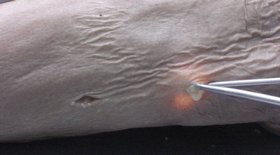
Fig. 3.2
Retrieval of the fascia lata using an optical dissector (Image courtesy of Mr. Stuart McNally.)
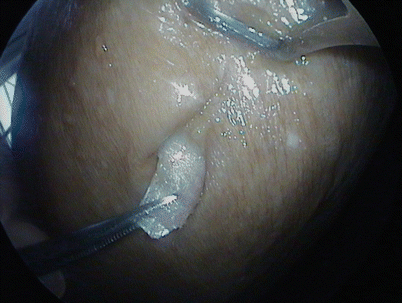
Fig. 3.3
Retrieval of the fascia lata strip through the proximal skin incision (Image courtesy of Mr. Stuart McNally.)
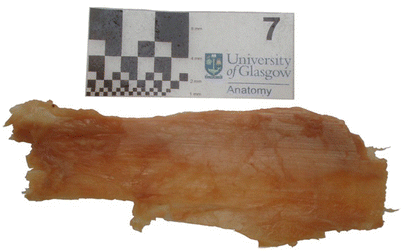
Fig. 3.4
Fascia lata taken from the cadaver in determining feasibility of endoscopic access for retrieval of the tissue. The scale bars include 1, 2, 4, and 8 mm and 1 cm. The maximum amount of fascia lata tissue retrieved during the experimental procedure was initially 90 × 30 mm (Image courtesy of Mr. Stuart McNally.)
Fascia lata is now commonly used to provide static facial support in cases of facial paralysis. When the facial nerve has little or no function, the mid face develops ptosis or sagging and the modiolus tends to droop. The result is an asymmetrical facial position at rest and oral incompetence, which cause great distress for the patient in regards to facial appearance and function. The affected side of the face can be lifted and supported by placing fascia lata slings under the skin to suspend the modiolus to a suitable support, usually the zygoma. Other indications for fascia lata grafts include revision of other surgical procedures used to reanimate the paralysed face, such as if a free muscle transfer has been used to “dynamise” a paralysed face but the position of the modiolus has slipped or was not sufficiently lifted. In these cases, a strip of fascia lata can be placed over the muscle to provide additional lift at the modiolus. Using an endoscopic approach for fascia lata harvest, strips of fascia up to 14 × 3 cm—a size easily sufficient for the technique of static facial slings—can be harvested from two small access incisions [19].
With no complications, reduced morbidity, and no scarring, it appears that the endoscopic approach to the fascia lata seems sensible to adopt into routine surgical procedures when access to this tissue is needed. For patients who are immunocompromised, elderly, or unable to undergo numerous operations, the use of autogenous fascia lata is ideal [4, 26]. With minimal scarring and no morbidity associated with endoscopic access, the ideal operative situation has been achieved [17–19].
3.2 Contraindications for the Use of Fascia Lata
The fascia lata should not be accessed for use in any reconstructive procedure if there has been previous trauma to the fascia lata or surgical retrieval of it. It is always pertinent to consider surgically accessing a site far away from the site that needs to be operated on. It may be easier to use temporalis fascia, especially on facial procedures like ptosis surgery. Surgery should not be performed until any infection at the site is satisfactorily treated [34].
3.3 Complications
Table 3.1 summarises the complications that can arise when traditional open access to the fascia lata is attempted, and lists relevant discussions of these pathologies. Endoscopic access to fascia lata, on the other hand, produces no major complications. The very slight prolongation of operating time is likely to disappear when surgical teams have more experience with endoscopic access to the fascia lata. With the lack of morbidity after the procedure, it makes sense to undertake the harvesting of the fascia lata by the endoscopic approach.
Table 3.1
Complications of open access to Fascia Lata
Complication | References |
|---|---|
Reduction in muscle power | Amir et al. [13] |
Visible scarring | Wheatcroft et al. [12] Amir et al. [13] Leibovitch et al. [2] Zandi [11] Kashkouli [6] |
Wound pain | Barron and Saad [32] Wheatcroft et al. [12] |
Limping/weakness of harvested side | Dubiel and Wigren [33] Wheatcroft et al. [12] |
Bleeding | Dubiel and Wigren [33] |
Wound dehiscence | Amir et al. [13] |
Muscle herniation | Dubiel and Wigren [33] |
3.4 Surgical Technique
The surgery is performed under general or regional anaesthesia with the patient in the supine position. For the harvest of fascia lata, a solution of local anaesthetic and adrenaline is infused around the upper, outer thigh in a volume depending on the amount of tissue to be harvested. The endoscope used to perform the procedure in all cases was the Karl Storz HOPKINS® ІІ Wide Angle Forward-Oblique Telescope (30°, 4 mm diameter, 18 cm length), and visual feedback was provided by a Karl-Storz Endoscope Tele Pack™ at 3× magnification.
Two 1- to 2-cm lazy-S skin incisions are made approximately 10 cm apart on the lateral aspect of the donor thigh. If the length of graft required is 5 cm or less, a single incision can be made. Using small retractors, dissection is made deeply to expose the fibres of the fascia lata, which should be clearly visualised as a tough, glistening layer in the base of the surgical field (Figs. 3.3, 3.5 and 3.6).
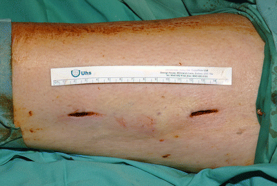
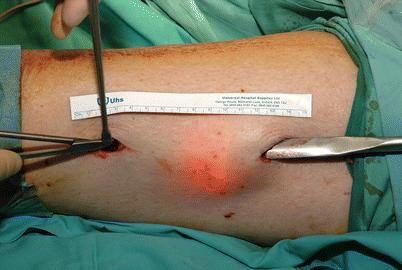
Fig. 3.5
Proximal (right) and distal (left) skin incisions carried out in the initial approach to access the fascia lata using the endoscopic technique in the operative environment
Fig. 3.6
Subcutaneous dissection over superficial aspect of the fascia lata, facilitated by an endoscope inserted into the proximal access incision
A subcutaneous pocket is developed over the fascia to be harvested. This plane is usually fairly avascular, but bipolar cautery is used if required. Through the upper skin incision, a 2- to 3-cm incision of the underlying fascia lata is made perpendicular to the skin. Passing under the fascia lata on the proximal side, it is separated from the underlying vastus lateralis by using an optical dissector attachment such as a Karl Storz TAKE-APART® elevator and Karl Storz CLICKLINE® scissors (Fig. 3.7). It is also separated from the overlying subcutaneous tissue by the same means. Using the endoscope for visualisation, the fascia lata is cut, ensuring a strip 2–3 cm wide all the way to the region of the distal skin incision for the desired length. The separated rectangular fascia lata strip, is then removed through the distal skin incision using fine forceps. Haemostasis is achieved using the endoscope and bipolar cautery.
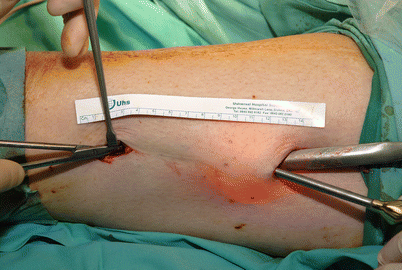
Fig. 3.7
Dissection proceeds using an endoscopic dissector in combination with scissors, with endoscopic assistance
The graft is defatted using scissors, and the recipient bed is prepared. If the graft is to be used as a primary static support, the length required is 12–13 cm. For revisionary procedures, a shorter length is often sufficient.
In the cheek, the local anaesthetic and adrenaline solution is infused subcutaneously. A pre-auricular, post-tragal incision is made and a tunnel is developed in the deep subcutaneous tissues. A small nasolabial incision of (1.5–2 cm) is made and the sub-cutaneous pocket is developed to communicate with this incision. If lip support is also required, the pocket is developed behind the vermillion of the upper and lower lip to the midpoint of the mouth or slightly beyond. Small incisions (5 mm) are made in the upper and lower lip.
The fascia is secured distally with sutures and fixed proximally to the zygomatic arch using a bone anchor (Fig. 3.8
 Video-Assisted Thoracoscopic Sympathicotomies for the Treatment of Palmar and Axillary Hyperhidrosis
 Minimally Invasive Endoscopic Surgical Treatment of Headache
 Endoscope-Assisted Brow Lift
 Transaxillary Endoscopic Subfascial Breast Augmentation
 Endoscopic Flap Design and Harvesting
 Breast Reconstruction Using Laparoscopically Harvested Omental Flap


Related posts:
Video-Assisted Thoracoscopic Sympathicotomies for the Treatment of Palmar and Axillary Hyperhidrosis
Minimally Invasive Endoscopic Surgical Treatment of Headache
Endoscope-Assisted Brow Lift
Transaxillary Endoscopic Subfascial Breast Augmentation
Endoscopic Flap Design and Harvesting
Breast Reconstruction Using Laparoscopically Harvested Omental Flap
Stay updated, free articles. Join our Telegram channel
Full access? Get Clinical Tree
