Fig. 7.1
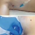
(a) A camera port and two to three 5-mm ports are inserted. Care must be taken not to injure the epigastric vessels, not only to avoid hemorrhagic complications but also to allow the potential for future total reconstruction. (b) The omentum is evaluated for size and adhesion. Then the patient is placed in reverse Trendelenburg position with a right lateral oblique rotation
First, the omentum is evaluated for size and adhesion (Fig. 7.1b) and is moved cephalic for dissection from the transverse colon.
The patient is placed in reverse Trendelenburg position with a right lateral oblique rotation. Usually a site slightly left of the center of the transverse colon is the most suitable place for starting dissection, providing easier access to the omental bursa (Fig. 7.2a).
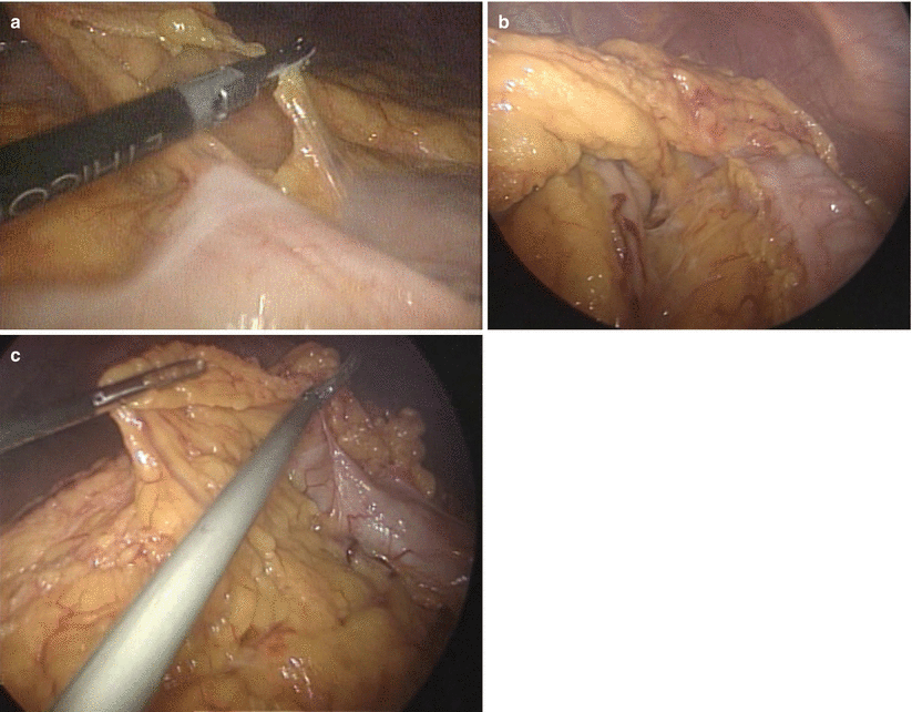
Fig. 7.2
(a) A site slightly left of the center of the transverse colon is the most suitable place for entering into the omental bursa. (b) To prevent injury of the omental flap, the posterior wall of the stomach must be identified in the first step of dissection. (c) Great care must be taken not to injure the transverse colon, not only by mechanical injury from grasping but also by thermal injury caused by the laparoscopic coagulating shears (LCS), especially near the splenic flexure
To ensure that the omental bursa is entered successfully, a posterior wall of the stomach must be identified (Fig. 7.2b).
This step is very important for safe and successful harvesting of the omental flap. Then the dissection is advanced leftward while maintaining appropriate tension between the omentum and the transverse colon. Care is taken not to injure the transverse colon, especially near the splenic flexure (Fig. 7.2c).
After the splenocolic ligament is divided, the resection turns to the lower pole of the spleen, with great care not to tear the splenic capsule (Fig. 7.3a).
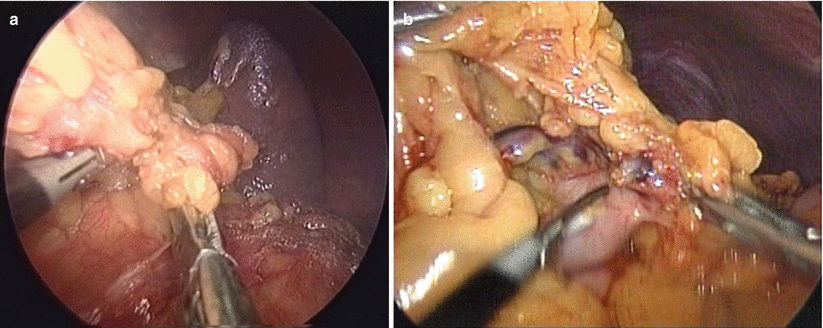
Fig. 7.3
(a) Transection of the left side of the omentum. After the splenocolic ligament is divided, the resection turns to the lower pole of the spleen, taking great care not to tear the splenic capsule. (b) The left gastroepiploic vessels can be resected with the LCS without ligation or clipping
During the resection of the gastrosplenic ligament, the left gastroepiploic artery and vein (GEAV) are encountered. They can be resected with a laparoscopic coagulating shears (LCS) without ligation or clipping (Fig. 7.3b).
Because the right gastroepiploic artery is predominant, we always select the right gastroepiploic artery and vein as a pedicle. The more volume of the flap is needed, the more resection of the gastrosplenic ligament beyond the short gastric vessels must be performed. However, the left side of the omentum beyond the resection wedge of the left gastroepiploic artery tends to lack adequate vascular supply.
After complete division of the left side of the omentum, the gastric branches of the GEAV are divided, little by little, at a site as close to the stomach wall as possible, towards the pyloric ring (Fig. 7.4a).
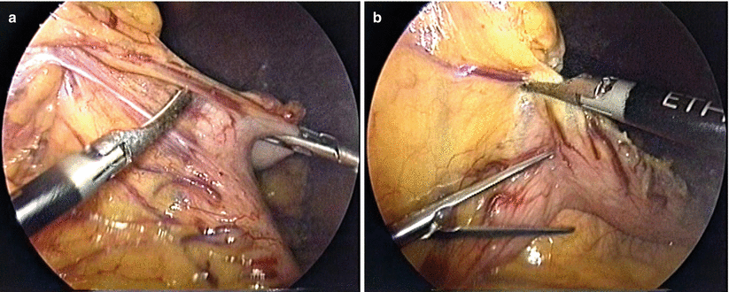
Fig. 7.4
(a) Unlike the transverse colon, the wall of the stomach is thick and strong. Then, the gastric branches of the gastroepiploic vessels should be divided at a site as close to the stomach wall as possible. (b) The gastric branches are pulled and stretched so as not to injure the main trunks of the gastroepiploic vessels
Attention is paid to the main trunks of the GEAV, because they sometimes run very close to the stomach wall, and they can be difficult to identify when abundant fat deposit of the omentum is evident. Therefore, the gastric branches of the GEAV are pulled and stretched so that they run at a right angle to the stomach wall (Fig. 7.4b).
The dissection from the right side of the transverse colon is then advanced (Fig. 7.5a), and fusion between the posterior leaf of the gastrocolic ligament and the anterior leaf of the transverse mesocolon is carefully divided toward the anterior capsule of the pancreas head (Fig. 7.5b).
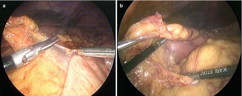
Fig. 7.5
(a) The dissection from the right side of the transverse colon is advanced with a left lateral oblique rotation. (b) Fusion between the posterior leaf of the gastrocolic ligament and the anterior leaf of the transverse mesocolon is bluntly divided with upward retraction of the stomach and downward retraction of the transverse mesocolon
Upward traction of the omentum is maintained, and careful sharp and blunt dissection between the gastrocolic ligament and the transverse mesocolon is advanced until the roots of the GEAV are confirmed. Adhesion to the anterior wall of the duodenum is also carefully dissected and advanced to the root of the GEAV.
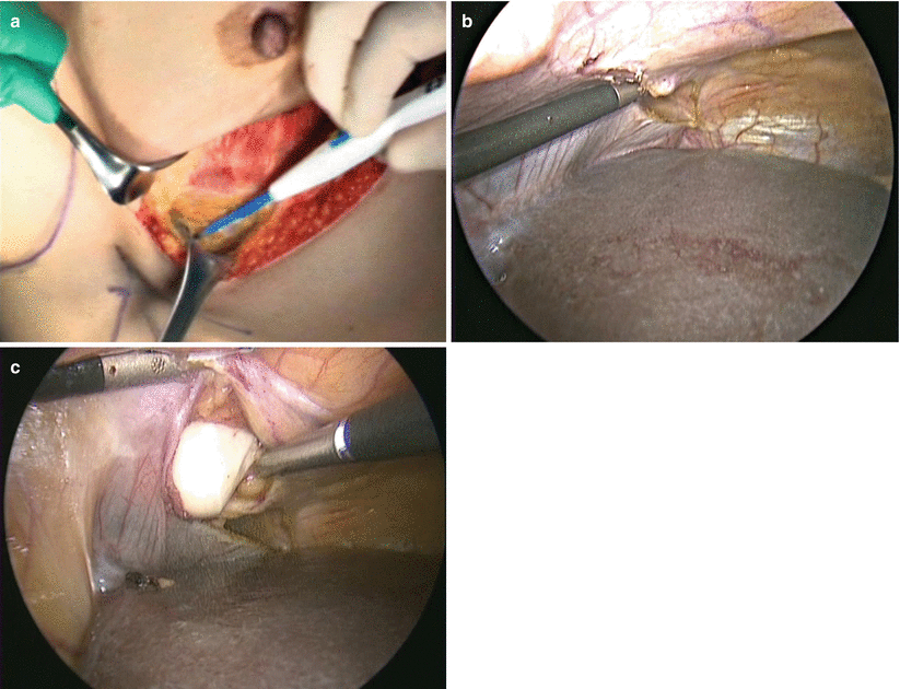
After complete dissection from the transverse colon, dissection from the stomach is advanced across the pyloric ring (Fig. 7.6a).
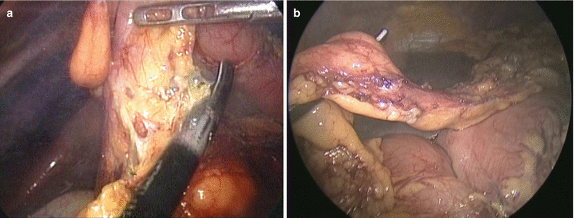
Fig. 7.6
(a) Resection of the gastric branches of the gastroepiploic vessels around the pyloric ring. (b) The root of the gastroepiploic vessels
Very careful dissection is required at this point, because the GEAV run close to the wall of the stomach and the proximal duodenum, and the branches tend to bleed easily. It is better to resect as much fat tissue around the root of the GEAV as possible for the thin pedicle of the flap, to avoid the subsequent complication of ventral hernia. One of the epiploic vessels that descends on the right side of the omentum sometimes must be resected to make the longer pedicle. Then a pedicled omental flap is completed (Fig. 7.6b).
7.3.2 Partial Breast Reconstruction After BCS
Using various skin incisions, the omental flap can be applied to any quadrants of partial mastectomy defects. For the lower quadrants, an inframammary skin incision is preferable (Fig. 7.7a, b).
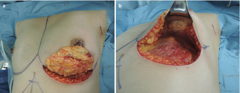
Fig. 7.7
(a, b) Partial mastectomy with an inframammary incision is the preferable choice for the omental flap to reconstruct a partial mastectomy defect in the lower quadrants
After partial mastectomy, a subcutaneous tunnel about two fingerbreadths in width is prepared from an incision along the inframammary fold toward the xiphoidal process (Fig. 7.8a). When it reaches the white line, a longitudinal incision measuring 2 or 2.5 fingerbreadths is made to communicate with the abdominal cavity.
 Endoscopic Harvest of the Fascia Lata for Facial Reanimation
 Video-Assisted Thoracoscopic Sympathicotomies for the Treatment of Palmar and Axillary Hyperhidrosis
 Endoscopic Diastasis Recti Repair
 Endoscope-Assisted Brow Lift
 Transaxillary Endoscopic Subfascial Breast Augmentation
 Endoscopic Flap Design and Harvesting


Related posts:
Endoscopic Harvest of the Fascia Lata for Facial Reanimation
Video-Assisted Thoracoscopic Sympathicotomies for the Treatment of Palmar and Axillary Hyperhidrosis
Endoscopic Diastasis Recti Repair
Endoscope-Assisted Brow Lift
Transaxillary Endoscopic Subfascial Breast Augmentation
Endoscopic Flap Design and Harvesting
Stay updated, free articles. Join our Telegram channel
Full access? Get Clinical Tree
