Fig. 2.1
With the patient supine and the head in a neutral position, frontal trigger nerves are located. Skin markings are drawn above the eyebrow bilaterally, at the mid-pupillary line (supraorbital nerve) and 1 cm medially (supratrochlear nerve)
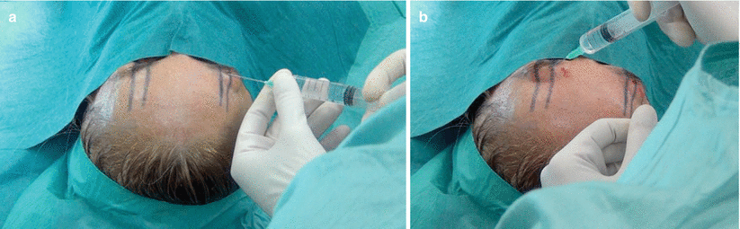
Fig. 2.2
The patient is put under sedo-analgesia and selective supratrochlear and supraorbital nerve block is performed on both on the right side (a) and the left side (b) in the glabellar region, following the skin markings. This block allows the injection of local anesthetic into the forehead to be completed in a painless manner
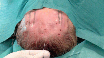
Fig. 2.3
Diluted carbocaine 1 % + 8.4 % sodium bicarbonate are infiltrated throughout the entire forehead, between the glabellar region and about 2 cm behind the anterior hairline. The local anesthetic has a twofold objective: not only anesthesia but also undermining of the tissues and creating a space between the periosteum and adjacent tissues to facilitate endoscopic visualization
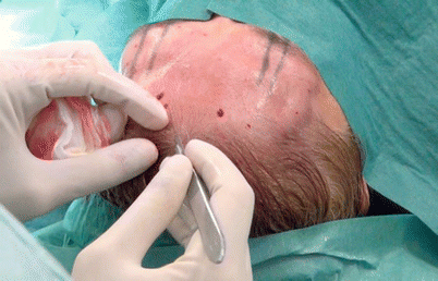
Fig. 2.4
A cutaneous incision 1.5 cm long is performed 1 cm behind the anterior hairline along the midline, dissecting all tissues (cutaneous, subcutaneous, aponeurotic galea) until periosteum is reached in the subgaleal plane. This location is chosen so that the postoperative scar will be hidden in the patient’s hair
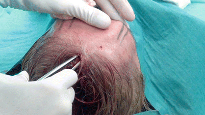
Fig. 2.5
Facilitated by the previous injection of local anesthetic, tissues all over the forehead bilaterally are undermined in the subgaleal plane through the hairline incision by means of long scissors. Undermining must be done carefully, particularly when the inferior limit of the undermining area (superciliary region) is reached, in order not to damage supratrochlear and supraorbital nerves. The lateral anatomic limit of the undermining area is the temporal region, bilaterally
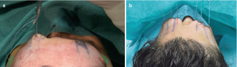
Fig. 2.6
(a, b), Nylon 1–0 sutures are placed in the superciliary region at each side of both supratrochlear and supraorbital nerves bilaterally, with the purpose of lifting the frontal skin during the endoscopic procedure and better visualizing the anatomic structures
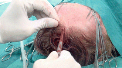
Fig. 2.7
To clean the subgaleal plane of blood and residual anesthetic fluid, suction of the entire undermined forehead is performed through the hairline incision before inserting the endoscope and whenever the endoscopic view is not clear
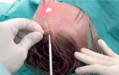
Fig. 2.8
The endoscope is inserted through the incision in the subgaleal plane until the superciliary region is reached, in order to perform the section of the corrugator supercilii, depressor supercilii, and procerus muscles bilaterally, thus decompressing the supraorbital and supratrochlear nerves bilaterally. Our modified endoscope (Karl Storz, Tuttlingen, Germany) consists of a 9-mm trocar with an air/insufflator/suction triple valve, a straight Hopkins telescope with fiber-light transmission, a Wittmöser operating sheath with a connection for high-frequency diathermy, and a specifically designed, elliptical-tipped wire loop electrode for electrocautery
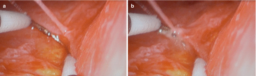
Fig. 2.9
The modified endoscope is used to perform endoscopically assisted section of the corrugator supercilii, depressor supercilii, and procerus muscles bilaterally, with the purpose of decompressing the supraorbital nerve, which is not injured during dissection. During this procedure, it is important to dissect every part of the muscle, which receives facial nerve fibers responsible for contraction of the muscle itself, in order to prevent irritation to surrounding nerves from the muscle’s movement. In these figures, the left supraorbital nerve is recognizable before (a) and during (b) the dissection; it is located in the superciliary region, 1 cm medial to the mid-pupillary line
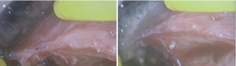
Fig. 2.10
Right supratrochlear nerve before (a) and during (b) the endoscopically assisted section of the right corrugator supercilii muscle. The supratrochlear nerve is located in the superciliary region, about 1.75 cm from the midline. Sections of glabellar muscles are performed at each side of both the supratrochlear and supraorbital nerves bilaterally. Endoscopic visualization is helpful in avoiding injury to the nerves and better identifying the muscles
Fig. 2.11
Surgical treatment of migraine and tension-type headache is also a useful procedure for occipital headaches, but headaches of this type require open surgery. In this patient, trigger sites have been identified in the greater occipital nerve (GON) bilaterally. Therefore, after performing local anesthesia, a horizontal occipital scalp incision 6 cm in length is performed along the superior nuchal midline to expose subcutaneous structures. No trichotomy is needed, and the scar from the incision will be hidden in the patient’s hair
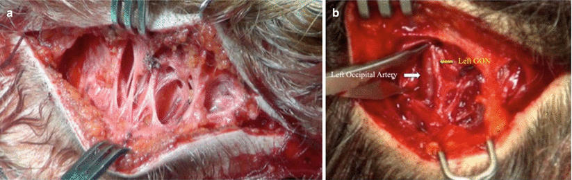
Fig. 2.12
 Endoscopic Harvest of the Fascia Lata for Facial Reanimation
 Video-Assisted Thoracoscopic Sympathicotomies for the Treatment of Palmar and Axillary Hyperhidrosis
 Endoscope-Assisted Brow Lift
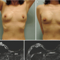
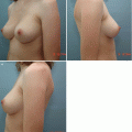 Transaxillary Endoscopic Subfascial Breast Augmentation
 Endoscopic Flap Design and Harvesting
 Breast Reconstruction Using Laparoscopically Harvested Omental Flap


Related posts:
Endoscopic Harvest of the Fascia Lata for Facial Reanimation
Video-Assisted Thoracoscopic Sympathicotomies for the Treatment of Palmar and Axillary Hyperhidrosis
Endoscope-Assisted Brow Lift
Transaxillary Endoscopic Subfascial Breast Augmentation
Endoscopic Flap Design and Harvesting
Breast Reconstruction Using Laparoscopically Harvested Omental Flap
Stay updated, free articles. Join our Telegram channel
Full access? Get Clinical Tree
