35 Wound Care
Quite often, patients will not want to or cannot see the wound. In this case, wound care may be demonstrated to a family member or the person who is accompanying the patient. A follow-up examination may be appropriate if the physician wants to assess the wound and/or the patient’s ability to care for it. Examples of wound care instructions are available in Appendix A.
Wound Healing Phases
Proliferative
The proliferative phase lasts from 2 days to 3 weeks. Macrophages, platelets, and fibroblasts release cytokines that initiate the formation of granulation tissue. Fibroblasts create a collagen bed to fill the defect and grow new capillaries. High lactate levels and low oxygen tension in the wound stimulate fibroblast proliferation and angiogenesis.1 Keratinocytes proliferate and migrate from the intact epidermis around the wound as well as from remaining structures in the base. The rate of re-epithelialization is directly related to moistness of the wound; open, dry superficial wounds re-epithelialize significantly more slowly than occluded moist wounds.2 One week after injury, myofibroblasts initiate contraction by pulling the wound edges closer together.
Remodeling
This phase lasts from 3 weeks to 2 years. Contraction continues, and an organized form of collagen gradually replaces the immature collagen. Wounds created by destructive techniques such as cryosurgery heal more slowly than those caused by a scalpel. Wound tensile strength is 40% at 1 month and never increases to more than 80% of the preinjury strength.1 This demonstrates the importance of supporting wounds with buried sutures and adhesive wound-closure strips, especially at the time of suture removal.
Types of Wounds
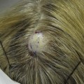
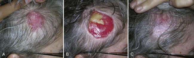
Two common types of skin wounds result from basic dermatologic surgery: a full-thickness wound that heals by primary intention and a partial-thickness wound that heals by secondary intention. However, even a full-thickness wound can be allowed to heal by secondary intention such as after a cancer removal on the scalp (Figure 35-1).
Dressings
Various types of dressings are available for use depending on the type of wound and physician preference. They are generally divided into open dressings (e.g., gauze) and occlusive dressings (e.g., films, foams, gels, hydrocolloids, and alginates). The selection and use of wound dressings can be highly personalized and modified by physician experience. The purpose of a dressing is to protect a wound from trauma or contamination, absorb wound drainage that may lead to maceration, provide hemostasis through compression, and facilitate healing by providing a moist environment. There is evidence that occlusive dressings increase re-epithelialization rates by 30% to 40% and collagen synthesis by 20% to 60% over air-exposed wounds.3 However, a randomized, controlled trial showed that there is no difference in infection between dressed and undressed clean sutured wounds.4 For physicians who do prefer to use wound dressings, characteristics required for the ideal dressing are found in Table 35-1.
TABLE 35-1 Ideal Dressing Characteristics
| Handling of excess exudate | Removal of toxic substances |
| Maintenance of moist environment | Barrier to microorganisms |
| Thermal insulation provided | Freedom from particulate contaminants |
| Removal without trauma to new tissue | Adheres well to a thin margin of surrounding skin |
| Does not adhere to wound | Nontoxic and nonreactive |
| Conforms well to body contours and motion | Promotes patient comfort and is not bulky |
| Readily available and inexpensive | Long shelf-life |
Source: From Freitag DS. Surgical wound dressings. In: Lask G, Moy R, eds. Principles and Techniques of Cutaneous Surgery. New York: McGraw-Hill; 1996.