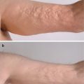
Fig. 11.1
Cleansing daily with soap and warm water should be encouraged for patients with sclerotic chronic GVHD. Before (a) and after (b); 2 days of soap and water cleansing followed by application of good emollient (100 % petrolatum)
Compression
Compression bandages and compression hosiery are forms of static compression therapy. Compression produces beneficial effects in wounds through multifactorial mechanisms. Traditionally, compression therapy has been most applied to the treatment of lower extremity venous stasis ulcerations, though similar principles may be useful in the treatment of GVHD wounds. Compression increases deep venous blood flow velocity and improves lymphatic flow and the local microcirculation. Compression therapy may also decrease the expression of pro-inflammatory matrix metalloproteinases, resulting in a wound milieu that favors ulcer healing [9]. Compression therapy is contraindicated in patients with peripheral artery disease, cellulitis, and acute deep vein thrombosis.
Compression bandages are generally divided into rigid (inelastic) and elastic types. The most common example of inelastic compression therapy is the Unna boot. Inelastic compression provides a high working pressure with muscle contraction during ambulation, but does not provide resting pressure. Elastic compression can be applied using elastic compression stockings, cotton/elastic wraps, short stretch bandages, or specialized multi-layered systems. Elastic compression bandaging systems conform to changes in leg size and sustain compression during activity and rest. Compression bandages should be applied by trained personnel and changed once or twice a week, depending on the degree of wound drainage [10].
Traditional compression stockings are often difficult for patients with sclerotic GVHD because of their restricted range of motion, neuropathic pain, skin fragility, and ulcer drainage. GVHD patients may respond better to multilayered wraps or combination therapy using specialized dressings directly placed on the ulcer bed that are then wrapped with compressive bandages.
Case 2
This patient, with extensive sclerotic chronic GVHD, was treated at a local wound center with serial debridement. At presentation, his legs demonstrated confluent sclerotic plaques with superficial and extensive erosions (Fig. 11.2a). He had poorly palpable pulses and pitting edema bilaterally. After starting a combination therapy including oral doxycycline, topical clindamycin and topical clobetasol ointment, he had significant improvement after 2 weeks (Fig. 11.2b). Although he had been given compression stockings by his hematologist and wound care physician, he could not apply them himself (due to his inability to bend at the hip or knees), and when placed by a caregiver the stockings would tear his fragile skin, worsening the erosions and causing additional pain. We recommended using cotton/elastic bandage wraps, which can be easily applied without causing additional trauma and can be adjusted to modify the amount of compression. Once his legs improved, we transitioned him to full length (foot, leg, and thigh) wraps secured with Velcro® straps that were much easier to apply and remove.
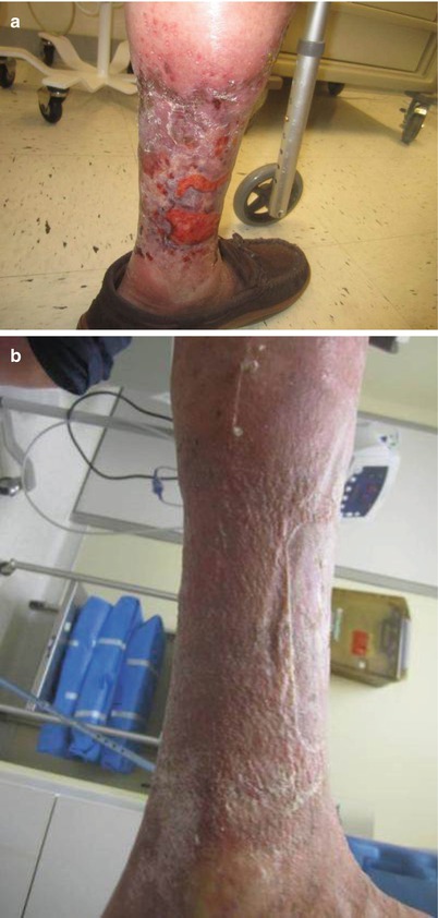
Fig. 11.2
(a, b) Surgical debridement is common in wound care centers but can lead to worsening chronic GVHD ulcers due to poor healing. A combination of topical steroid and compression was used for this patient, who demonstrated significant and lasting improvement
Topical and Intralesional Steroids
Topical Steroids
Topical corticosteroids are an appropriate first-line therapy for patients with mild cutaneous disease. As skin atrophy is known to be a major side-effect of long-term treatment, care should be taken in prolonged use, particularly for the face and intertriginous areas of the body. However, for many of these patients, the benefit of decreasing systemic immunosuppression far outweighs the risk of epidermal atrophy. Systemic absorption of topical steroids may be a concern in pediatric patients, but in these patients benefits also generally outweigh these risks [11]. Treatment should start with high-potency topical steroids. In unresponsive patients, short-term occlusion with damp dressings increases skin hydration and steroid penetration.
Topical treatment of mild GVHD is important especially for patients with a high risk of relapse, as increasing systemic immunosuppression may interfere with the desired graft-versus-malignancy effect [11]. In moderate-to-severe GVHD, topical immunosuppression may be used in conjunction with systemic immunosuppression to increase local response rates. Furthermore, topical immunosuppressive therapies for cGVHD are associated with less toxicity compared with systemic treatment, and may allow for dose reduction of systemic immunosuppression [11].
Intralesional Steroids
Intralesional steroids may be helpful for localized cutaneous GHVD refractory to high-dose topical steroid therapy. Mid-potency steroids such as triamcinolone 1 mg/kg is often used, and often requires multiple treatments. While effective in clinical practice, its use is often limited by a lack of consensus guidelines and patient discomfort associated with the procedure. However, in patients who have refractory localized disease, intralesional injections can resolve their lesions and allow them to discontinue systemic immunosuppression.
Case 3
A 10-year-old male patient had extensive sclerotic cGVHD affecting 60–70 % BSA. He developed chronic ulcerations of his scalp that were resistant to systemic immunosuppression. At presentation, he had numerous crusted erosions localized to the scalp (Fig. 11.3, left panel). Numerous topical therapies were attempted without significant improvement, including antibiotics, steroids, and calcineurin inhibitors. After extensive discussion of the risks and benefits, we elected for a trial of intralesional steroids. The patient required sedation for the procedure and a total of 1 mg/kg was injected into four trial areas. After significant improvement was noted in these lesions, the patient underwent three additional treatments using 1–2 mg/kg of intralesional triamcinolone at each treatment. He had complete resolution of his lesions (Fig. 11.3, right panel) and was weaned completely off systemic immunosuppression. Of note, he also exhibited partial reversal of his alopecia.
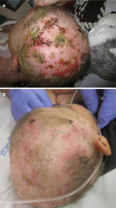
Fig. 11.3
(a, b) When ulcers are localized, intralesional steroids can be used to heal chronic ulcers and decrease systemic immunosuppression
Topical Calcineurin Inhibitors
For patients who fail topical or intralesional therapy, topical calcineurin inhibitors (CNIs) may be considered. Topical CNIs (e.g., tacrolimus and pimecrolimus) are useful localized steroid-sparing medications. However, most patients who initially respond to topical calcineurin inhibitors eventually require additional treatment. Topical CNIs may be of particular benefit for sites at high risk of skin atrophy (lips, eyelids, and intertriginous surfaces). CNIs are generally poorly tolerated at areas of very active skin involvement with erosions [12].
Complex Wounds
Complex, chronic wounds, such as those associated with cGVHD, contain a persistently high amount of inflammatory exudate, an environment that impedes the proliferation of fibroblasts [13]. Local therapy plays an important role in diminishing skin inflammation and promoting wound healing. Ideal dressings are those that control exudate, prevent bacterial proliferation, and absorb excessive wound drainage while preventing drying.
Related posts:
 Grading and Treatment of Acute Graft-Versus-Host Disease
 Diagnosis, Staging, and Treatment of Chronic Graft-Versus-Host Disease
 Clinical Presentation of Nonsclerotic Epidermal Chronic Graft-Versus-Host Disease and Hair and Nail Changes
 Dermal and Subcutaneous Chronic Graft-Versus-Host Disease
 Diagnosis, Staging, and Treatment of Chronic Graft-Versus-Host Disease
 Clinical Presentation of Nonsclerotic Epidermal Chronic Graft-Versus-Host Disease and Hair and Nail Changes
Stay updated, free articles. Join our Telegram channel

Full access? Get Clinical Tree


