Fig. 11.1
(a) Arm volume meter. Figures in millimeter. (b) The clinical setup
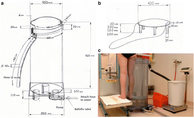
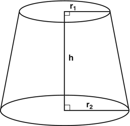
Fig. 11.2
(a) Leg volume meter. Figures in millimeter. (b) Leg volume meter. Spacers to compensate for various leg lengths. Figures in millimeter. (c) The clinical setup
For both the arm and leg volume meter it is important that the draining pipe is be placed high enough so that the entire limb can be measured. The diameter of the draining hose should be large so that the water can flow quickly. It is important that the limb is immersed into the volume meter at exactly the same depth each time.
Arm Volume Meter (Fig. 11.1)
The hose is connected to a standard faucet with a threaded bayonet mount and the volume meter is filled with water through a hose at the bottom of tank. The arm is brought down into the volume meter with the palm against the wall to avoid surge. The fingers are held outstretched and brought down until the tip of the middle finger reaches the bottom. Wait a few seconds to allow the water to flow out. There are gradation lines with 1-cm intervals for patients who do not reach down to the bottom of the volume meter. Measurements can be reproduced from time to time by noting the distance between the volume meter’s bottom and the tip of the middle finger. The drained water is collected in a plastic tray (Orth Plast, for example, model 101-8: 40 cm × 34 cm × 16 cm), which holds about 17 L. To empty the volume meter the hose is led down a drain in the floor or to a sink so the water can drain. An electric pump accelerates the runoff (Eheim 1046, Eheim, http://www.eheimparts.com). A drawing of a volume meter is shown in Fig. 11.1. The scale should have an accuracy of 5 g (digital max 30 kg/5 g). The volume meter has an average measurement error (CV%) of 0.24.
Leg Volume Meter (Fig. 11.2)
To compensate for different leg lengths, different spacers are put into the volume meter so that the entire leg volume can be measured up to the crotch. The measuring cylinder’s height minus the leg length = the height of the spacer. An electric pump accelerates the runoff when emptying (Eheim 1060, Eheim, http://www.eheimparts.com). To hoist the patient a commercially available “lift” (Santo Lift SC 200, PMH International AB, http://www.pmh.se) is used. The water is collected in a large plastic laundry basket (Idealplast; model 0519; 33 cm × 55 cm), which holds about 40 L (Fig. 11.2).
Volume Measurements Based on Circumference Measurements
Another method is circumference measurements on well-defined distances along the limb. Circumference measurements also provide information of the localization of the swelling. Hand and foot volumes cannot be calculated. To only make single circumference measurements (for example the middle of the upper arm, elbow, middle of the forearm) is not adequate either for clinical use or scientific study.
Volume measurements based on plethysmography or circumference measurements are useful and show satisfactory validity and reliability [4–9], plethysmography is recommended if only one method is used [10].
How Volumes Are Calculated Using Circumference Measurements
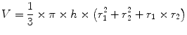
The circumferences of the limb measured every 4 cm along the limb. The volume of each segment is calculated according to the formula of the truncated and added together to get the limb volume (Fig. 11.3):
 You need:
Fig. 11.3
Truncated cone segment. H height of the cone. r 1 radius at upper end of the segment. r 2 radius at lower end of the segment
A long ruler to mark every the 4 cm
Marker pen (eyeliner pen)
Narrow tape measure for circumference measurements
Protocol.
How to measure:
Arm lymphedema: The patient sits on a chair with the arm straight and abducted 90 ° on a table
Highlight the 0 point in the wrist level
The tape is placed around the extremity distal to each mark
Read the circumferences measured and record
Leg lymphedema: The patient sits with the hip flexed at 90° with extended knee and foot in 0-position.
The measurement programs for arms and legs can be downloaded from: https://lu.box.com/s/9abvigfbx2rw7afgk9kz or by contacting the first author.
Other Methods
The following methods are not used routinely, but mainly for research purposes.
Perometry
This is an optoelectronic measuring method using a square frame with multiple perpendicular light beams. The frame is moved along the limb and the cross-sectional area is calculated continuously and thereby the volume. The equipment is expensive, but the method is accurate and fast [12]. Foot and hand volume are not measured [13, 14].
Bioelectric Impedance (BIS)
Local Tissue Fluid, Tissue Dielectric Constant (TDC)
Tissue fluid may be measured by means of electromagnetic waves transmitted via a measuring head, which is set against the skin. The instrument measures a few millimeters into the skin and a constant (tissue dielectric constant), directly proportional to the tissue fluid content, is calculated. The method is useful for measuring tissue water anywhere in the body where the skin is not too close to the skeleton. The method has been used to measure edema in the extremities [17], legs [18, 19].
Volume Calculations
A large number of computational methods have been presented for reporting treatment results [5, 7, 8]. The excess volume of a unilateral lymphedema is calculated as the difference between the edematous and the contralateral healthy extremity.
The absolute excess volume (volume of the affected limb − volume of the unaffected limb) of a lymphedema [22, 23] together with the relative value in percent (affected limb/unaffected limb × 100) gives optimal information before and after treatment. The relative value provides information about the extent of the edema [1, 10]. The same absolute excess volume provides greater relative value (swollen extremity/healthy extremity) in a skinny person as compared to a corpulent [1, 10].
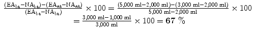
Absolute Excess Volume and Percentage Reduction
Edematous arm (EA), Normal arm (NA), Before treatment (bt), After treatment (at).
EA | NA | Excess volume | Excess volume reduction (%) | |
|---|---|---|---|---|
bt | 5,000 ml | 2,000 ml | ∆3,000 ml | |
67 % | ||||
at | 3,000 ml | 2,000 ml | ∆1,000 ml |
By measuring the normal arm at the same time as the edematous arm one takes into account the daily variations in arm volumes as well as weight gain or loss. It is very important that this is done at every time when volumes are measured.
 Follow-up controls
EA | NA | Excess volume | % Excess volume reduction | |
|---|---|---|---|---|
bt | 5,000 ml | 2,000 ml | ∆3,000 ml | |
at1 | 4,000 ml | 2,200 ml |


