Primary (prevalence 1/1,00,000 persons)
Secondary (prevalence 1/1,000 persons)
Infant-onset
Filariasis
Adolescent-onset
Axillary/inguinal lymphadenectomy
Childhood-onset
Axillary/inguinal radiation
Adult-onset
Obesity
Primary Lymphedema
Primary lymphedema is rare, affecting approximately 1/100,000 children [5]. In the pediatric population onset occurs in infancy (49.2 %), childhood (9.5 %), or adolescence (41.3 %) [1]. Primary lymphedema develops during adulthood in 19 % of patients [6]. Males are more likely to present in infancy (68 %), while females most commonly develop the disease during adolescence (55 %) [1]. The lower extremities are affected in 91.7 % of patients; 50 % have unilateral lymphedema and 50 % have bilateral disease [1]. Bilateral lower extremity lymphedema is more common in patients presenting in infancy (63 %), compared to adolescence (30 %) [1]. Eighteen percent of children with primary lymphedema have genital disease, which is usually associated with lower extremity lymphedema. Four percent of patients with primary lymphedema have isolated genital involvement. Sixteen percent of children with idiopathic lymphedema have upper extremity disease [1]. Rarely, a child can have lymphedema affecting the legs, genitalia, and/or arms.
Secondary Lymphedema
Cancer-Related Treatment
Injury to the lymphatic system is responsible for approximately 99 % of adult cases and 3 % of pediatric disease [1]. The overall risk of lymphedema following treatment for malignancy is 15 %; the two variables that most importantly predict if a patient develops the condition is if he/she underwent lymphadenectomy or radiation [7]. The overall risk of lymphedema after treatment for the following cancers (including patients who did and did not have lymphadenectomy or radiation) has been estimated to be: head/neck (4 %), genitourinary (10 %), melanoma 16 % (arm 5 %, leg 28 %), gynecologic (20 %), sarcoma (30 %) [7].
Upper extremity lymphedema from breast cancer treatment is the most common etiology of the disease in the USA. One-third of women who have axillary lymphadenopathy and radiation develop the condition [8]. Edema typically begins 12 months following the injury to lymph vessels [9]. Three-fourths of patients develop swelling within 3 years after the injury and the risk of lymphedema is 1 % each year thereafter [10]. Advanced disease, the extent of resection, and number of lymph nodes removed increases the risk of lymphedema [11]. Modified radical mastectomy has a greater chance of causing lymphedema compared to partial mastectomy; removal of more than 15 axillary lymph nodes increases the rate of lymphedema tenfold, compared to excising less than 5 nodes [12]. Sentinel lymph node biopsy reduces the rate of lymphedema (0.5 %) compared to axillary lymph node dissection [13]. Radiation is a major risk factor for breast cancer-related lymphedema; when the axilla is included in the radiation field the risk of lymphedema doubles, compared to radiation of the breast and supraclavicular nodes only [14]. In patients who have undergone lymphadenectomy and/or radiation, the most significant variable that will increase their risk of lymphedema is obesity [15].
Pelvic or abdominal malignancy is the most frequent reason for lower extremity lymphedema. The rate of lymphedema following lymphadenectomy and/or radiation for the following malignancies has been estimated to be: prostate (13 %), uterine (18 %), melanoma (25 %), vulvar (28 %), sarcoma (25 %). penile (30 %), cervical (42 %) [4]. Lower-extremity and genital lymphedema rates decrease when inguinal sentinel lymph node biopsy is performed (1.9 %) instead of lymphadenectomy [16].
Filariasis
A parasitic infection is the most common etiology of lymphedema in the world; 90 % of cases are cause by W. bancrofti [2]. Eighty-three countries are endemic to the disease; 70 % of cases are in Bangladesh, India, Indonesia, and Nigeria [2]. Other affected areas include Africa (central), Brazil, Burma, China (southern), Dominican Republic, Guiana, Guyana, Haiti, Malaysia, Nile delta, Pacific Islands, Pakistan, Sri Lanka, Surinam, and Thailand [2]. It is estimated that although 120 million people are infected with a lymphedema-causing parasite, 40 million persons exhibit lymphedema clinically [2]. Patients at risk for filariasis live in tropical/subtropical environments because these habitats are humid which is necessary for the parasites to survive [2]. Most individuals with filariasis live in rural locations that have poor sanitation [2]. The most common location for lymphedema caused by filariasis is the lower extremity and/or genitalia, but the upper extremity and breast can be affected as well.
Obesity
Obesity affects one-third of the US population, and 6 % have a body mass index (BMI) >40 [17]. The proportion of the population that is obese is increasing at a rate of 2–4 % every 10 years [17]. Patients with a BMI > 50 are at risk for developing obesity-induced lower extremity lymphedema, and individuals with a BMI > 60 are very likely to have the disease. Although the percent of the US population with a BMI > 60 is unknown, using a conservative estimate of 0.5 % then the number of Americans (population 315 million) with obesity-induced lymphedema would be 1.575 million.
Morbidity
Some patients with lymphedema do not have problems, while others can have significant complications (Table 4.2). Generally, individuals who are compliant with intervention have less disability than patients who are noncompliant (Fig. 4.1). Individuals with an active lifestyle have fewer problems than patients who are sedentary. Exercise likely improves proximal lymphatic flow by muscle contraction and helps the individual maintain a normal BMI. Obese individuals have more complications from lymphedema compared to normal-weighted persons because obesity adversely affects lymphatic function [15, 18]. Morbidity from lymphedema is described below from the most common problem, to the least frequent complication.
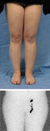
Table 4.2
Morbidity from lymphedema (listed from most common to least common)
Progression of disease |
Lowered self-esteem |
Infection |
Fitting clothing |
Difficulty using the extremity |
Skin changes |
Massive localized lymphedema |
Discomfort |
Malignant transformation |
Fig. 4.1
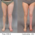
Asymptomatic lymphedema. 19-year-old female with recent swelling of the right lower extremity. Lymphoscintigram shows absent transit of tracer to the right inguinal nodes 2 h following injection. The patient does not have any complaints and the appearance of her legs is similar
Progression
Almost all patients with lymphedema have progression of their disease. Over time the high-protein interstitial fluid causes subcutaneous adipose deposition [19]. Consequently, the circumference of the limb or genitalia gradually enlarges. Fat in an extremity can increase by 73 % [19]. As the lymphedematous area becomes larger, the patient’s ability to use the area decreases.
Psychosocial
Lymphedema negatively impacts a patient’s social well-being, body image, and sexuality [20]. The most common problem expressed by patients with lymphedema is that they are unhappy with the appearance of their limb or genitalia. Patients have lowered self-esteem because the involved area does not look normal (Fig. 4.2). The more severe the disease, the greater the loss of self-esteem and negative impact on quality of life. Unilateral limb involvement can be more distressing than bilateral disease because the asymmetry is more noticeable. Individuals may not feel comfortable wearing clothing that exposes their diseased limb. Patients may avoid changing clothes in front of their peers or refrain from swimming. Lymphedema can impede the establishment of new relationships and genital disease can negatively impact an individual’s sexual activity.
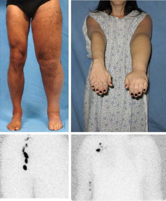
Fig. 4.2
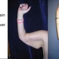
Psychosocial morbidity caused by lymphedema. (Left) 50-year-old male with adult-onset left-lower extremity lymphedema. (Right) 40-year-old female with left upper extremity lymphedema following breast cancer treatment. Both patients had lowered self-esteem because they were unhappy with the appearance of their diseased limb
Patients with secondary lymphedema from cancer treatment often state that although they are cured of their malignancy, lymphedema is a daily reminder of their cancer. Children with primary disease often feel that they are “different” than their peers; the emotional aspect of their condition worsens during adolescence. Many patients believe that wearing a compression garment is a worse deformity than the appearance of their lymphedematous extremity. The most common reason for individuals to seek surgical intervention is to improve the appearance of the area affected by lymphedema. Patients with psychosocial distress can be helped with counseling. Conservative compression strategies, as well as excision of excess subcutaneous fat using liposuction, can improve a patient’s asymmetry as well as their self-esteem.
Infection
The most frequent “functional” problem caused by lymphedema is infection. A lymphedematous extremity has a significantly increased risk of cellulitis compared to the non-affected limb (Fig. 4.3). Lymph stagnation increases the risk of infection after minor trauma because of: (1) impaired immunosurveillance (lymphatics function as an immunologic defense), (2) decreased oxygen delivery to the skin, and (3) a proteinaceous environment favorable for bacterial growth. In one community study of both primary and secondary lymphedema, 29 % of patients had an infection over the previous 12 months, and one-fourth required hospitalization for intravenous antibiotics [3]. In another study of patients with primary lymphedema, 19 % have a history of cellulitis, 13 % have been hospitalized, and 7 % have >3 attacks each year [1].
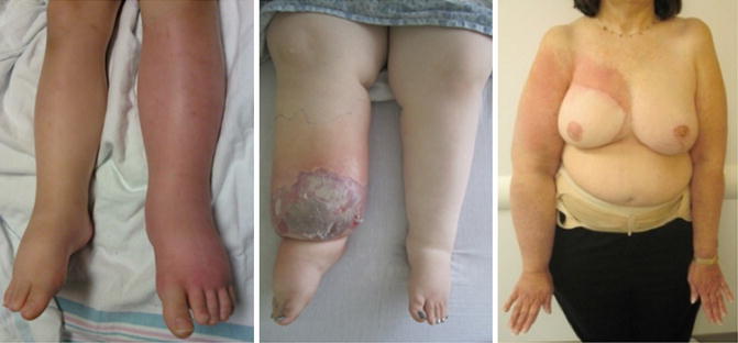
Fig. 4.3
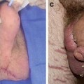
Lymphedematous areas are at increased risk for infection. (Left) 4-year-old male with primary lymphedema developed left leg cellulitis requiring hospital admission. (Center) 18-year-old female with primary lymphedema had right lower extremity infection necessitating intravenous antibiotics; note skin epidermolysis and blistering. (Right) 64-year-old female with cellulitis complicating secondary lymphedema of the right arm following breast cancer treatment
Infections in a lymphedematous area typically do not spontaneously occur, but result from a break in the skin. The most common etiology is incidental trauma; the patient is unaware of a cut or scrape. Less frequently, the source of an infection can be a problem with a finger/toe nail. Rarely, a systemic infection can secondarily infect a lymphedematous extremity.
Related posts:

Stay updated, free articles. Join our Telegram channel

Full access? Get Clinical Tree


