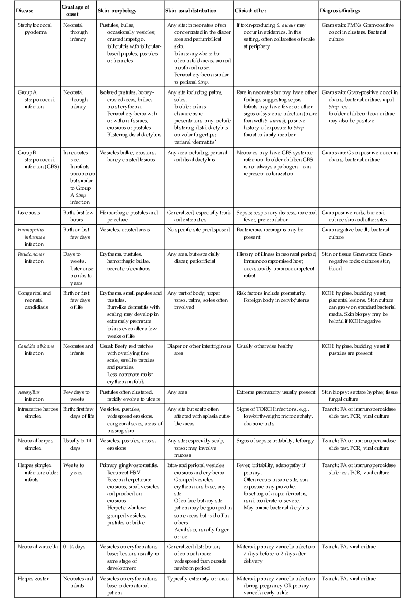
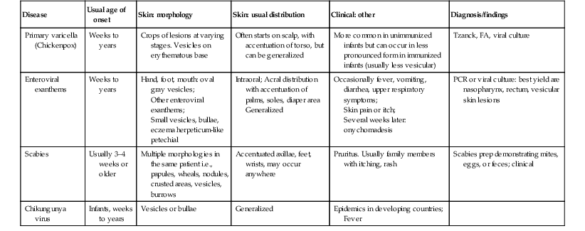
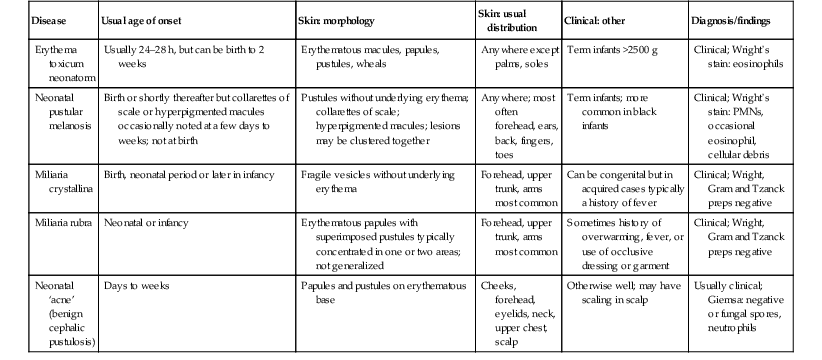
Vesicles, Pustules, Bullae, Erosions, and Ulcerations


10

Vesicles, Pustules, Bullae, Erosions, and Ulcerations


Renee M. Howard, Ilona J. Frieden


Introduction


Vesiculopustular and bullous disorders are common in the neonatal period and the first years of life. Accurate and prompt diagnosis is essential because some conditions that present with blisters and pustules are truly life-threatening. In contrast, many others are innocuous and self-limited; misdiagnosis of a more serious condition can lead to iatrogenic complications, unnecessary expense, and parental anguish.


The causes of blisters and pustules in newborns and young infants are influenced by the clinical setting, including geography and whether patients are seen in a hospital or clinic. Infection is the most common etiology in developing countries. In a study of neonates in India with blisters and pustules, bacterial infection was the most common overall, whereas erythema toxicum was the most prevalent non-infectious etiology.1 In a prospective study of newborn inpatients in California examined by pediatric dermatologists, the most common eruption in newborns was erythema toxicum.2 In a retrospective European study done in a Level 3 nursery, vesicles and pustules were the sole reason for admission to the Neonatal Intensive Care Unit (NICU) in 29.8% of infants admitted because of skin lesions.3


Several articles have reviewed an approach to infants with these cutaneous findings,46 including infants with hemorrhagic vesiculopustules.7 There is also considerable overlap with the subject matter in other chapters of this book, most notably Chapter 7 (Transient Benign Cutaneous Lesions in the Newborn), and Chapters 12, 13 and 14 (Bacterial, Viral, and Fungal Infections, respectively), so the main discussion of specific disorders are discussed therein. Chapter 11 discusses the diagnosis and management of epidermolysis bullosa and other non-infectious causes of bullae, so those conditions are covered in far less detail in this chapter.


In addition to a discussion of vesicles, pustules and bullae, this chapter also includes conditions presenting with erosions and ulcerations. Although vesicles, pustules, and bullae are primary skin lesions, they can quickly progress to secondary skin lesions (i.e. erosions and ulcerations). This can occur rapidly or have transpired in utero, such that erosions and ulcerations are the main presenting finding. Examples include staphylococcal scalded skin syndrome, where erythema and skin erosions predominate over blisters, and Pseudomonas skin infection, where pustules rapidly evolve into necrotic ulcers.


Because of the wide range of diagnoses discussed in this chapter, there are boxes and tables to help with a systematic approach to evaluation and differential diagnosis. Tables 10.110.3 summarize key findings and differential diagnosis of vesiculopustular diseases in newborns and infants, including infectious causes, relatively common transient skin lesions, and uncommon and rare causes of this clinical presentation. Tables 10.410.6 summarize these same categories for the differential diagnosis of bullae, erosions, and ulcerations. Box 10.1 lists conditions in neonates where pustules and vesicles predominate, Box 10.2 lists the conditions in neonates where bullae predominate, and Box 10.3 lists conditions in neonates where erosions or ulcerations may predominate.


TABLE 10.1


Differential diagnosis of vesiculopustular diseases in newborns and infants – Infectious causes


Disease Usual age of onset Skin: morphology Skin: usual distribution Clinical: other Diagnosis/findings
Staphylococcal pyoderma Neonatal through infancy Pustules, bullae, occasionally vesicles; crusted impetigo, folliculitis with follicular-based papules, pustules or furuncles Any site: in neonates often concentrated in the diaper area and periumbilical skin.
Infants: anywhere but often in fold areas, around mouth and nose.
Perianal erythema similar to perianal Strep.
If toxin-producing S. aureus may occur in epidemics. In this setting, often collarettes of scale at periphery Gram stain: PMNs Gram-positive cocci in clusters. Bacterial culture
Group A streptococcal infection Neonatal through infancy Isolated pustules, honey-crusted areas, bullae, moist erythema.
Perianal erythema with or without fissures, erosions or pustules.
Blistering distal dactylitis
Any site including palms, soles.
In older infants characteristic presentations may include blistering distal dactylitis on volar fingertips; perianal ‘dermatitis’
Rare in neonates but may have other findings suggesting sepsis.
Infants may have fever or other signs of systemic infection (more than with S. aureus), positive history of exposure to Strep. throat in family member
Gram stain: Gram-positive cocci in chains; bacterial culture, rapid Strep. test.
In older children throat culture may also be positive
Group B streptococcal infection (GBS) In neonates – rare.
In infants uncommon but similar to Group A Strep. infection
Vesicles bullae, erosions, honey-crusted lesions Any area including perianal and distal dactylitis Neonates may have GBS systemic infection. In older children GBS is not always a pathogen – can represent colonization Gram stain: Gram-positive cocci in chains; bacterial culture
Listeriosis Birth, first few hours Hemorrhagic pustules and petechiae Generalized, especially trunk and extremities Sepsis; respiratory distress; maternal fever, preterm labor Gram-positive rods; bacterial culture skin and other sites
Haemophilus influenzae infection Birth or first few days Vesicles, crusted areas No specific site predisposed Bacteremia, meningitis may be present Gram-negative bacilli; bacterial culture
Pseudomonas infection Days to weeks.
Later onset months to years
Erythema, pustules, hemorrhagic bullae, necrotic ulcerations Any area, but especially diaper, periorificial History of illness in neonatal period, Immunocompromised host; occasionally immunocompetent infant Skin or tissue Gram stain: Gram-negative rods; cultures skin, blood
Congenital and neonatal candidiasis Birth or first few days of life Erythema, small papules and pustules.
Burn-like dermatitis with scaling may develop in extremely premature infants even after a few weeks of life
Any part of body; upper torso, palms, soles often involved Risk factors include prematurity.
Foreign body in cervix/uterus
KOH: hyphae, budding yeast; placental lesions. Skin culture can grow on standard bacterial media. Skin biopsy may be helpful if KOH negative
Candida albicans infection Neonates and infants Usual: Beefy red patches with overlying fine scale, satellite papules and pustules.
Less common: moist erythema in folds
Diaper or other intertriginous area Usually otherwise healthy KOH: hyphae, budding yeast if pustules are present
Aspergillus infection Few days to weeks Pustules often clustered, rapidly evolve to ulcers Any area Extreme prematurity usually present Skin biopsy: septate hyphae; tissue fungal culture
Intrauterine herpes simplex Birth; first few days of life Vesicles, pustules, widespread erosions, congenital scars, areas of missing skin Any site but scalp often affected with aplasia cutis-like areas Signs of TORCH infections, e.g., low-birthweight; microcephaly, chorioretinitis Tzanck; FA or immunoperoxidase slide test, PCR, viral culture
Neonatal herpes simplex Usually 5–14 days Vesicles, pustules, crusts, erosions Any site; especially scalp, torso; may involve mucosa Signs of sepsis; irritability, lethargy Tzanck; FA or immunoperoxidase slide test, PCR, viral culture
Herpes simplex infection: older infants Weeks to years Primary gingivostomatitis.
Recurrent HSV
Eczema herpeticum: erosions, small vesicles and punched-out erosions
Herpetic whitlow: grouped vesicles, pustules or bullae
Intra- and perioral vesicles erosions and erythema
Grouped vesicles erythematous base, any site
Often face but any site – pattern may be grouped in some areas but trail off in others
Acral skin, usually finger or toe
Fever, irritability, adenopathy if primary.
Often recurs in same site, sun exposure may provoke.
In setting of atopic dermatitis, usual moderate to severe.
May mimic bacterial dactylitis
Tzanck; FA or immunoperoxidase slide test, PCR, viral culture
Neonatal varicella 0–14 days Vesicles on erythematous base; Lesions usually in same stage of development Generalized distribution, often much more widespread than outside newborn period Maternal primary varicella infection 7 days before to 2 days after delivery Tzanck, FA, viral culture
Herpes zoster Neonates and infants Vesicles on erythematous base in dermatomal pattern Typically extremity or torso Maternal primary varicella infection during pregnancy OR primary varicella early in life Tzanck, FA, viral culture
Primary varicella (Chickenpox) Weeks to years Crops of lesions at varying stages. Vesicles on erythematous base Often starts on scalp, with accentuation of torso, but can be generalized More common in unimmunized infants but can occur in less pronounced form in immunized infants (usually less vesicular) Tzanck, FA, viral culture
Enteroviral exanthems Weeks to years Hand, foot, mouth: oval gray vesicles;
Other enteroviral exanthems;
Small vesicles, bullae, eczema herpeticum-like
petechial
Intraoral; Acral distribution with accentuation of palms, soles, diaper area
Generalized
Occasionally fever, vomiting, diarrhea, upper respiratory symptoms;
Skin pain or itch;
Several weeks later: onychomadesis
PCR or viral culture: best yield are nasopharynx, rectum, vesicular skin lesions
Scabies Usually 3–4 weeks or older Multiple morphologies in the same patient i.e., papules, wheals, nodules, crusted areas, vesicles, burrows Accentuated axillae, feet, wrists, may occur anywhere Pruritus. Usually family members with itching, rash Scabies prep demonstrating mites, eggs, or feces; clinical
Chikungunya virus Infants, weeks to years Vesicles or bullae Generalized Epidemics in developing countries;
Fever


imageimage


TABLE 10.2


Differential diagnosis of vesiculopustular diseases – Transient skin lesions


Disease Usual age of onset Skin: morphology Skin: usual distribution Clinical: other Diagnosis/findings
Erythema toxicum neonatorm Usually 24–28 h, but can be birth to 2 weeks Erythematous macules, papules, pustules, wheals Anywhere except palms, soles Term infants >2500 g Clinical; Wright’s stain: eosinophils
Neonatal pustular melanosis Birth or shortly thereafter but collarettes of scale or hyperpigmented macules occasionally noted at a few days to weeks; not at birth Pustules without underlying erythema; collarettes of scale; hyperpigmented macules; lesions may be clustered together Anywhere; most often forehead, ears, back, fingers, toes Term infants; more common in black infants Clinical; Wright’s stain: PMNs, occasional eosinophil, cellular debris
Miliaria crystallina Birth, neonatal period or later in infancy Fragile vesicles without underlying erythema Forehead, upper trunk, arms most common Can be congenital but in acquired cases typically a history of fever Clinical; Wright, Gram and Tzanck preps negative
Miliaria rubra Neonatal or infancy Erythematous papules with superimposed pustules typically concentrated in one or two areas; not generalized Forehead, upper trunk, arms most common Sometimes history of overwarming, fever, or use of occlusive dressing or garment Clinical; Wright, Gram and Tzanck preps negative
Neonatal ‘acne’ (benign cephalic pustulosis) Days to weeks Papules and pustules on erythematous base Cheeks, forehead, eyelids, neck, upper chest, scalp Otherwise well; may have scaling in scalp Usually clinical; Giemsa: negative or fungal spores, neutrophils


image


TABLE 10.3


Differential diagnosis of vesiculopustular diseases – Uncommon and rare causes


Only gold members can continue reading. Log In or Register to continue

Stay updated, free articles. Join our Telegram channel

Apr 27, 2016 | Posted by in Dermatology | Comments Off on Vesicles, Pustules, Bullae, Erosions, and Ulcerations

Full access? Get Clinical Tree

Get Clinical Tree app for offline access
Disease Usual age of onset Skin: morphology Skin: usual distribution Clinical: other Diagnosis/findings
Acropustulosis of infancy Birth or days to weeks Vesicles and pustules Hands and feet, occasional lesion elsewhere Severe pruritus accompanying lesions which tend to come in crops Clinical; skin biopsy: intraepidermal vesicle/pustule
Eosinophilic pustular folliculitis Birth or days to weeks Pustules, erythema Mainly scalp and face; occasionally trunk, extremities Pruritus; waxing and waning course with recurrent crops Skin biopsy: dense perifollicular mixed infiltrate with eosinophils
Incontinentia pigmenti Birth to days Vesicles, hyperkeratosis in linear array Most common on trunk, scalp, extremities Extracutaneous involvement common but often not evident at birth.
Mothers may have history of IP or other findings (e.g., missing teeth, areas of decreased hair growth)
Skin biopsy: eosinophilic spongiosis with dyskeratosis
Gene testing can also be used to establish diagnosis in atypical cases
Neonatal Behçet disease First week of life Small punched out vesicles, pustules, ulcerations and scarring Perioral and oral mucous membranes, hands and feet, occasionally other sites Maternal history of Behçet disease Clinical findings and maternal history
Erosive pustular dermatosis of the scalp Weeks to months Crusting, pustules, scaly erythema Scalp, superimposed on areas of alopecia, scarring from scalp injury Severe scalp edema or necrosis of delivery; similar findings in Hay–Wells and Rapp–Hodgkin ectodermal dysplasias Clinical findings and prior history of scalp injury or ectodermal dysplasia
Hyper-IgE syndrome Days to months Single or grouped pustules, vesicles, or crusting Face, scalp, upper torso Blood eosinophilia
Note: IgE levels often become elevated after neonatal period
Skin biopsy: intraepidermal vesicle with eosinophils or eosinophilic folliculitis.
Gene testing for STAT-3 mutation
Lipoid proteinosis Usually ≥1 year Erythematous papulovesicular lesions resulting in atrophic scarring Face, ears, extremities and occasionally trunk Thickening of the skin, especially lips, perinasal skin, tongue; hoarseness Skin biopsy shows thick hyalinized material with characteristic PAS-positive staining.
Positive FH in some cases
Pustular psoriasis or deficiency of interleukin-1 receptor antagonist First weeks or months of life Pustules generalized, but especially palms, soles; may have underlying erythroderma Generalized Irritability, occasionally fever Skin biopsy: epidermal microabscesses and acanthosis, parakeratosis, dilated capillaries
Pustular eruption of myelodysplasia in Down syndrome/neonatal eosinophilic pustulosis First few days to months of life Extensive pustules on erythematous base, often aggregating in areas of skin injury Face most common site but can occur elsewhere Very high WBC count: usually in setting of Down syndrome but can occur without obvious Down phenotype or with other causes of severe leukocytosis