, Shimin Chang2, Jian Lin3 and Dajiang Song1
(1)
Department of Orthopedic Surgery, Changzheng Hospital Second Military Medical University, Shanghai, China
(2)
Department of Orthopedic Surgery, Yangpu Hospital Tongji University School of Medicine, Shanghai, China
(3)
Department of Microsurgery, Xinhu Hospital Shanghai Jiao Tong University, Shanghai, China
The anatomical basis and the clinical application of the vastus medialis perforator flap was firstly reported by Zheng and Lin [1].
20.1 Vascular Anatomy
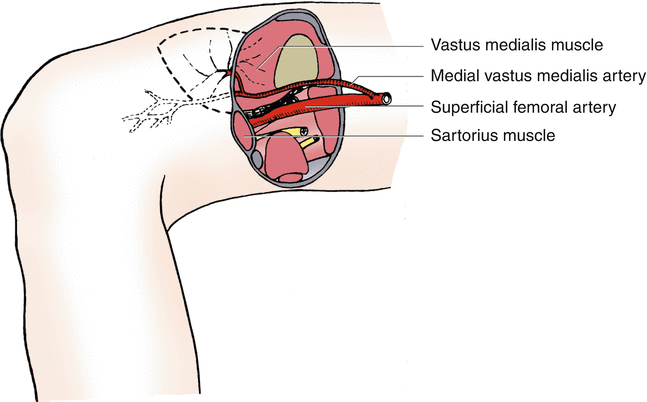
A large muscular artery, named the medial vastus medialis artery, is given off from the superficial femoral artery at the tip of the femoral triangle, which then travels laterally downward along the muscle fibers after entering the muscle and anastomoses with the rete patellae when it reaches the knee joint. The site of the first musculocutaneous perforator is relatively constant, whose piercing point into the fascia can be located around the midpoint of the surface projection line (a line drawn from the junction of the middle and lower thirds of the line between the midpoint of the inguinal groove and the medial femoral condyle to the midpoint of the upper margin of the patella) of the medial vastus medialis artery (Fig. 20.1).


Fig. 20.1
Vascular anatomy of the medial vastus medialis artery
20.2 Illustrative Case
A 28-year-old female sustained a soft tissue defect at the anterior aspect of the right knee in a traffic accident.
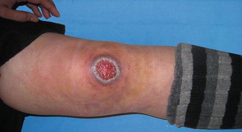
During operation, the wound was radically debrided at first, resulting in a defect measuring 5.0 ×3.3 cm (Fig. 20.2).


Fig. 20.2
Preoperative view
Flap Design
Stay updated, free articles. Join our Telegram channel

Full access? Get Clinical Tree


