Vascularized Rib-Periosteal Transplantation for Reconstruction of The Mandible and Maxilla
D. SERAFIN
V. E. VOCI
EDITORIAL COMMENT
This technique has now been supplanted by free fibular/scapular osteocutaneous flaps and iliac-crest flaps.
Nonvascularized bone grafts have had reasonable success in small, well-vascularized defects. The major problem arises when larger defects require large segments of contoured bone and soft-tissue coverage. The rib is one source of vascularized bone that can be transplanted with or without overlying skin (1, 2).
INDICATIONS
Vascularized rib-periosteal transplantation is indicated to reconstruct a moderate deficiency of the maxilla or mandible in a relatively avascular bed. The recipient skin envelope must be adequate to accommodate the relative bulk of the osseous segment and associated bulky musculature.
If the skin envelope is healthy with adequate blood supply and the mandibular or maxillary defect is small to moderate in size, then a nonvascularized bone graft will provide satisfactory results. If the mandibular or maxillary segment to be replaced is large with an associated large cutaneous defect, then a vascularized iliac osteocutaneous flap based on the deep circumflex iliac vessel is the donor tissue of choice (3).
The major advantage of the vascularized rib-periosteal transplant is the ability to provide vascularized osseous tissue in a relatively avascular bed. Osteocyte survival has been confirmed by subsequent biopsies, arteriography, and serial radiographs (4, 5, 6). lone union occurs with callus formation similar to that noted at a fracture site.
ANATOMY
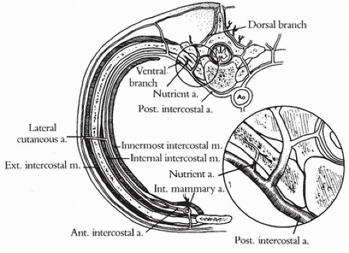
There are nine paired posterior intercostal arteries. They arise from the dorsal aspect of the aorta. At their origin, they lie midway in the intercostal space and course obliquely upward as they proceed laterally. The arteries on the right are longer because the aorta lies to the left of the vertebral column. Opposite the heads of the ribs, the posterior intercostal artery divides into a ventral and dorsal branch (Fig. 205.1). The dorsal branch supplies the spinal cord, its meninges, and the vertebrae. It then continues dorsally to supply muscular branches to the paraspinous muscles and cutaneous branches to the skin. The artery of Adamkiewicz arises from one of these dorsal branches between T7 and L2. This is an extremely important vessel because it provides the major blood supply to the thoracolumbar spinal cord. Injury to this vessel may result in paraplegia.
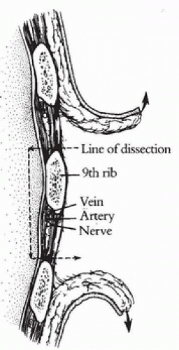
Proximally, the ventral branch of the posterior intercostal artery lies between the posterior intercostal membrane and pleura. By the time it has reached the angle of the rib, however, it has penetrated the posterior intercostal membrane and internal intercostal muscle and continues anteriorly between the external and internal intercostal muscles. It courses upward in the interspace obliquely to lie inferior to the corresponding rib and continues anteriorly in the costal groove between the pleura and the posterior intercostal membrane. It eventually anastomoses with the anterior intercostal artery, a branch of the internal thoracic artery, at about the midclavicular line. Each intercostal artery is accompanied by a vein and an intercostal nerve. The vein is superior and the nerve is inferior to the artery (Fig. 205.2).
The ventral branch gives off multiple branches. The first branch is the nutrient artery to the rib above. This nutrient branch is given off near the tubercle of the rib and enters the bone through a nutrient foramen (see Fig. 205.1). It quickly divides into proximal and distal medullary arteries in the medullary cavity of the rib. Collateral intercostal branches are given off at regular intervals, beginning at the angle of the rib. They course down to the superior border of the rib below, supplying the surrounding soft tissue. Musculocutaneous perforators are also given off at regular intervals, supplying the pectoralis, serratus, and intercostal muscles as well as the overlying skin. The overlying skin is also supplied by a lateral cutaneous arterial branch that arises at the midaxillary line and courses with the lateral cutaneous nerve. The direct and musculocutaneous perforators supply the skin portion of an osteocutaneous flap. Mammary branches are given off by the third, fourth, and fifth intercostal arteries.
The veins on the right side empty into the azygous vein. On the left side, the upper veins empty into the accessory hemiazygous vein, and the lower veins empty into the hemiazygous vein.
OPERATIVE TECHNIQUE
Three operative approaches have been described for rib-periosteal and osteocutaneous rib grafts: anterior, posterolateral, and posterior. Each method has advantages and disadvantages.
 FIGURE 205.2 A cross section of the rib and the corresponding interspaces. Note the location of the intercostal vein, artery, and nerve. |


