Type of VCA model
Specific feature
Skin and soft tissue containing VCA models
Groin flap as vascularized skin transplant model
Semimembranosus muscle and epigastric skin transplant model
Total abdominal wall transplant model
Hindlimb model
Rat hind limb transplant model
Groin-thigh osteomyocutaneous transplant model
Immunomodulatory VCA models
Bone marrow transplantation
Thymus transplantation
Facial allograft models
Models containing partial or total facial structures, such as face, hemiface or hemiface mandible
Models including functional unit
Other VCA model
Models of certain organs and tissues, such as penis, larynx, lymph node
Table 19.2
Basic experimental models of VCA and their specific features
Experimental model | Specific features of model | |
|---|---|---|
Skin containing VCA models | Groin flap as vascularized skin transplant model | Simple, used in immunologic studies, small skin component |
Semimembranosus muscle and epigastric skin transplant model | Used for microcirculatory, physiologic, and immunologic studies | |
Total abdominal wall transplant model | Contains large skin component | |
Hindlimb models | Rat hind limb transplant model | Popular, but technically challenging animal model containing all tissue components including vascularized bone marrow |
Groin-thigh osteomyocutaneous transplant model | Alternative model to hind limb transplant, less challenging technique, low mortality | |
Immunomodulatory VCA models | Vascularized femur transplant model | Used for bone marrow induced chimerism studies |
Bilateral vascularized femoral bone transplant model | Larger bone marrow content, applied for tolerance induction studies | |
Composite vascularized skin/bone transplant model | Combination of groin flap with vascularized femur transplantation, enables follow-up of rejection | |
Vascularized osteomyocutaneous iliac transplant model | Includes both vascularized bone marrow and large skin amount | |
Vascularized sternum transplant model | Includes sternum, skin and muscles, contains vascularized bone marrow | |
Thymus transplant model | Used for evaluation of effect of thymus on tolerance induction | |
Osseomusculocutaneous sternum, ribs, thymus, pectoralis muscles, and skin transplant | Includes all three important immunologic tissues (bone marrow, thymus and skin) for immunology and tolerance studies | |
Facial allograft models | Full face/scalp transplant model | Includes ear, neck, and facial skin without eyelids and nose, high mortality |
Hemiface/scalp transplant model | Unilateral ear, neck, and facial skin. Used for immunologic studies in facial transplantation | |
Hemiface calvaria transplant model | Temporal muscle and parietal bone included to hemiface scalp model | |
Hemiface mandible tongue allotransplantation model | Mandibular bone and tongue is included into hemiface model. Contains bone marrow | |
Maxilla transplant model | Used for evaluating maxillary response to transplantation | |
Midface transplant model with sensory and motor units | Includes functional unit. First model to evaluate motor and sensory recovery after face transplantation | |
Total osteocutaneous hemiface transplant model | Includes entire hemiface, ideal model for immunological, histological and biological evaluation of different facial tissues | |
Composite face and eyeball transplant model | Includes functional unit. Evaluation of optic nerve regeneration and evaluation of orbit content |
Skin and Soft Tissue-Containing VCA Models
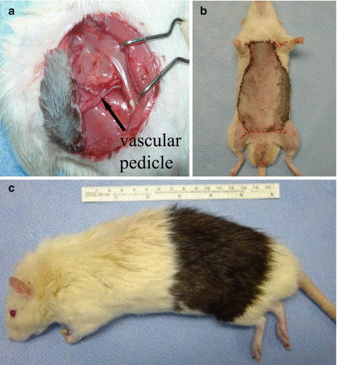
Skin and soft tissue-containing VCA models include allografts containing skin, subcutaneous tissue, fat and muscle (Fig. 19.1). The most frequently used skin/soft tissue VCA model is the groin flap, which is also one of the first models used in VCA research [7]. This model contains skin, fat and lymphoid tissue harvested on superficial epigastric vessels or femoral vessels. The groin flap is preferred for immunologic studies as it is technically simple and transplantation procedures have low mortality rates. The main disadvantage of this model is the small size of the skin paddle. Later, various VCA models including, skin, subcutaneous tissue and different muscles (such as semimembranosus and pectoralis) were described [8, 9]. Another soft tissue model is the total abdominal wall transplantation model in which the entire abdominal wall skin is harvested on bilateral femoral vessels to investigate the effect of skin amounts on chimerism [10].


Fig. 19.1
Examples of skin and soft tissue-containing VCA models. (a) Groin flap, (b) total abdominal wall flap, (c) extended groin flap
Hindlimb Allotransplantation Models
The first rat hindlimb allotransplantation model was described in 1978 [3]. In this model, a circumferential skin incision at the mid-thigh level is made and femoral vessels, saphenous nerve, sciatic nerve and femoral nerve are dissected and thigh muscles are sharply cut. Next, the femoral bone is cut with a saw. The recipient is prepared using the same technique and the donor hindlimb is transferred to the recipient. Following the fixation of the femoral bone with an intramedullary rod and wire suture, femoral vessels are subsequently anastomosed and the femoral, sciatic and saphenous nerves are coaptated. Muscles and skin are closed anatomically. This transplantation is a complex procedure and quite similar to human hand transplantation. However, it is time consuming, requires expertise and has a high mortality rate. Thus, simpler models are needed to obtain good survival of the recipient animal and robust statistics. Later, Chang et al. described transplantation of the groin-thigh osteomyocutaneous flap, which is composed of skin (groin), muscle (thigh), and bone (two-thirds of the femur), based on the femoral vessels and superficial epigastric vessels [11]. The advantages of this model are shortened operative time, easy monitoring of graft rejection and decreased mortality rates.
Immunomodulatory VCA Models
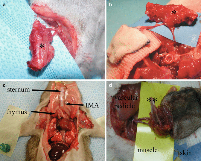
Immunomodulatory VCA models include immunologic tissues such as bone marrow and thymus, to increase chimerism and induce immunotolerance (Fig. 19.2). Generally, the most frequently used tissue for immunomodulation is bone marrow, especially femoral bone marrow. Tai et al. described a vascularized femur transplant comprising femoral bone, bone marrow, periosteum, cartilage and muscle tissues [12]. This model was used to investigate the effects of vascularized bone marrow transplantation on chimerism induction. Later, a bilateral, vascularized femoral bone transplantation model was described by Agaoglu and Siemionow [13]. This model included the same tissue types, but with a higher bone marrow content, which induced higher levels of donor-specific chimerism. Another immunomodulatory model is the composite vascularized skin/bone allograft model, which is basically a combination of the groin flap model with vascularized femur transplantation [14]. The skin component helps to follow-up the rejection and bone marrow is used to induce chimerism. The osteomyocutaneous iliac flap is composed of iliac bone, abdominal wall musculature, and a wide skin island, harvested on iliolumbar artery and vein [15]. The sternum is also used as a bone marrow source in VCA studies. The sternum, including presternal skin and muscles, was first transplanted by Santiago et al. [16]. The allograft was harvested on the thoracic aorta and superior vena cava.


Fig. 19.2
Examples of immunomodulatory VCA models. (a) Vascularized femoral flap (*, femoral bone), (b) vascularized femoral bone and groin flap, (c) vascularized osteomusculocutaneous sternum, thymus allograft, (IMA internal mammary artery), (d) vascularized iliac bone-skin allograft (**, iliac bone)
When the amount of bone marrow cells is compared, it is seen that the limb model contains 48.75 × 10⁶ bone marrow cells. On the other hand, iliac, femur, and sternum models contain 25, 50, and 7.5 × 10⁶ bone marrow cells, respectively.
Another method of tolerance induction in experimental studies is to include thymus tissue. The first thymus transplantation was described by Jiang et al. in 1999 [17]. Later, composite osseomusculocutaneous sternum, ribs, thymus, pectoralis muscles, and skin allotransplantation models were developed [18].
Facial VCA Models
The face has different anatomic units and structures. Thus, there are many facial VCA models based on the transplantation of these structures (Fig. 19.3). Siemionow et al. were the first to develop a full-face scalp allotransplantation model [4


Stay updated, free articles. Join our Telegram channel

Full access? Get Clinical Tree


