Fig. 24.1
Schematic presentation of vascularized thymus transplantation. (a) Donor. The right lobe of the thymus on its vascular pedicle, including the subclavian artery and superior caval vein, is harvested. (b) Recipient. Thymus is placed in the right side of the recipient’s neck and is anastomosed to the common carotid artery and to the jugular vein
The arterial supply of the right thymic lobe originates from the brachiocephalic artery and branches out into subclavian artery, internal thoracic artery, and finally the thymic artery. The venous drainage from the right lobe originates from the thymic vein and drains into the internal thoracic, subclavian, and superior caval veins. All irrelevant side branches are ligated to create a single vascular pedicle. The right lobe of the thymus, after all thymus-associated lymph nodes are removed, is separated from the left lobe. The subclavian artery is clamped just distal to the bifurcation and sharply transected. The superior caval vein also sharply cut horizontally just proximal to the right atrium. The right lobe of the thymus on its vascular pedicle, including both the subclavian artery and superior caval vein, is harvested and placed in the right side of the recipient’s neck prepared previously. The subclavian artery of the harvested thymus is anastomosed to the recipient’s common carotid artery, and superior caval vein to the jugular vein, using conventional end-to-end microsurgical with 9/0 sutures, respectively. The transplanted right lobe of the thymus is positioned within the neck cavity created by removal of the sternal head of sternocleidomastoid muscle. The skin is closed with 5/0 Vicryl suture.
Evaluation of Viability of Vascular Thymus Transplant
Immediately following transplantation, perfusion of the transplanted thymus is evaluated by two different microsurgical patency tests at 5 and 30 min after anastomosis: (a) the milking, and (b) the hook patency tests.
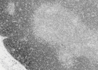
The flow of the anastomosed subclavian artery and superior caval vein are evaluated with a Model T-206 flowmeter (Transonic). Histologic H&E sectioning and examination is done at the end of the planned experimental study (Fig. 24.2).


Fig. 24.2
Histologic sections of vascularized thymus transplant demonstrating normal architecture of the thymus with preserved lobes and germinal centers
The reason for transplantation failure could be attributed to vascular anatomic variations, severe vasospasm, patchy appearance, and venous congestion due to the ‘no-outflow’ phenomenon, as observed in Sprague-Dawley rats, as well as air embolization and kinking. Subcapsular hematoma and subsequent fibrosis coul be attributed as the late complications.
Stay updated, free articles. Join our Telegram channel

Full access? Get Clinical Tree


