Classification of vascular lesions based of off the biological behavior has greatly facilitated more accurate diagnoses, optimally defined treatment plans, and better outcomes. Treatment of vascular lesions has taken a more conservative surgical approach with reliance on select medical treatment options, which has greatly reduced morbidity and mortality resulting from extensive surgery. A multidisciplinary approach involving multiple surgical and pediatric subspecialties has led to advancement in both understanding and ideal treatment strategies of these lesions.
Key points
- •
Classification of vascular lesions based of off the biological behavior has greatly facilitated more accurate diagnoses, optimally defined treatment plans, and better outcomes.
- •
Treatment of vascular lesions has taken a more conservative surgical approach with reliance on select medical treatment options, which has greatly reduced morbidity and mortality resulting from extensive surgery.
- •
A multidisciplinary approach involving multiple surgical and pediatric subspecialties has led to advancement in both understanding and ideal treatment strategies of these lesions.
Introduction
The study of vascular lesions has spanned many medical and surgical subspecialties within academia over the last several decades. Much of the understanding regarding the nonsurgical management of these lesions has been predicated by pediatric subspecialties and complemented by advances in surgical management of these lesions and vice versa. Furthermore, because many of these lesions have involved the craniofacial region, this area has been thoroughly investigated by surgical subspecialties, including facial plastic surgery along with oral maxillofacial and craniofacial surgery.
Since the advent of the current classification system approved by the International Society for the Study of Vascular Anomalies in 1996 (and updated in 2014), relatively few advances in treatment of these lesions have occurred. This classification system was derived from Mulliken and Glowacki’s system, which was published in 1982 and based on the biological behavior of the lesions. In general, the overlying theme has been toward more conservative surgical approaches and a greater reliance on the advances in medical management, which in turn has led to more effective treatment with a greatly reduced morbidity.
Specifically, the lesions can be divided into vascular tumors, including the more common infantile hemangiomas, rapidly and noninvoluting congenital hemangiomas, and kaposiform hemangioendotheliomas, among others; and vascular malformations, including low-flow venous, lymphatic, and capillary/port-wine stain subtypes, high-flow arteriovenous malformations (AVM), and combined complex capillary-venous, capillary-arteriovenous, and lymphaticovenous subtypes. In this article, the more common lesions, and specifically the classification, diagnosis, and management, are focused on, with an emphasis on an aesthetic approach regarding surgical technique when indicated ( Box 1 ).
Vascular tumors
- •
Infantile hemangiomas
- •
Congenital hemangiomas
- ○
Rapidly involuting
- ○
Noninvoluting
- ○
Partially involuting
- ○
- •
Kaposiform hemangioendothelioma
- •
Others
Vascular malformations
- •
Low-flow
- ○
Venous malformations
- ○
Lymphatic malformations
- ○
Capillary/port-wine stain malformations
- ○
- •
High-flow
- ○
Arteriovenous malformations
- ○
- •
Combined complex
- ○
Capillary-venous
- ○
Capillary-arteriovenous
- ○
Lymphaticovenous
- ○
Vascular tumors: hemangioma
Considered the most common tumor of infancy and childhood, hemangiomas have an estimated prevalence of 10% by age 1, with 30% evident at birth, and a female predilection of 4:1. Ninety percent of all vascular tumors are infantile hemangiomas. Although 40% to 60% involve the head and neck, 80% of these lesions tend to be solitary. There are various syndromes that can be associated with hemangiomas, including PHACES ( p osterior fossa malformations, h emangiomas, a rterial anomalies, c oarctation of the aorta and cardiac defects, e ye abnormalities, and s ternal defects), and other symptoms such as stridor in a patient with cutaneous hemangiomas should be further investigated along with the potential for visceral hemangiomas and hemangiomas overlying the lumbosacral spine due to the increased association with spinal cord and genitourinary anomalies. Imaging studies, such as screening ultrasounds and confirmatory MRIs, should be strongly considered in these patients.
Historically, treatment of these lesions involved the use of various modalities. Many investigators advocated for aggressive management with surgical excision, cauterization, carbon dioxide snow, surface radium, radioactive implants, external beam radiation, interstitial gamma radiation, and sclerosing agents. However, serious complications arose, such as malignant transformation after radiation therapy and poor cosmetic results due to significant scar formation secondary to resection and other ablative procedures. Conversely, proponents of more conservative benign neglect strategies argued that the natural course of most hemangiomas included involution with no residual deformity. The stark polarity in treatment algorithms was centered around the fact that vascular tumors and malformations were often categorized as similar or related entities—for example, the term capillary hemangioma was used to describe port-wine stains, which are capillary malformations, whereas strawberry nevi and cavernous hemangiomas were used to describe true hemangiomas. This early misclassification of vascular lesions is what led to a lack of consensus in regards to management, which again was addressed by Mulliken and Glowacki’s delineation of vascular tumors (hemangiomas) and malformations based on biological behavior, resulting in the current classification.
Clinical presentation and diagnosis
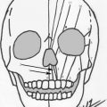
Infantile hemangiomas are usually visible within the first few weeks of life, whereas congenital hemangiomas are fully formed at birth and grow with variable intensity. Superficial hemangiomas are contained within the papillary dermis and are characterized by bright red, macular, or papular lesions with well-defined borders. The macular variety can be confused with port-wine malformations; however, over time the hemangioma will change in size, whereas the port-wine malformation remains relatively stable. Deep hemangiomas are contained in the reticular dermis or subcutaneous tissue and present as a blue, subcutaneous mass with bluish or colorless overlying skin depending on the depth. These hemangiomas may appear similar to lymphatic malformations on examination. Compound hemangiomas have features of both superficial and deep lesions ( Fig. 1 ).
Hemangiomas exhibit 2 distinct clinical stages, including proliferation and involution. Proliferation occurs during the first 12 months of life and occasionally as late as 18 months. An initial growth phase during the first few months of life followed by a subsequent growth phase at 4 to 6 months establishes a bimodal pattern of growth. Cosmetic deformity or functional obstruction of the eye, nose, or airway typically arises during the first growth phase ( Fig. 2 ). Histologic features during the proliferative phase include plump proliferating endothelial cells and pericytes with barely perceptible vascular channels. Involution, occurring over the first several years of life, is characterized by a decrease and then cessation in growth of the lesion. The lesion changes from bright red to dark maroon and eventually patches of ashen gray, evolving from a firm, tense consistency to a lobular, soft, compressible mass on palpation. Histologically, there is a gradual flattening of endothelial cells with progressive deposition of fibrous tissue and vessel ectasia, resulting in superficial telangiectasia and subcutaneous fibrofatty residuum ( Fig. 3 ).
Although imaging is not required for diagnosis, it can be useful to evaluate the extent of involvement in deeper lesions as well as rule out other vascular tumors. Ultrasound findings demonstrate a solid mass with increased/high color flow within it; Doppler ultrasound can demonstrate the arterial feeder and venous drainage. MRI characteristics include lesions that exhibit enhancement on T2-weighted images, which are relatively isointense on T1-weighted images with homogenous contrast enhancement; internal serpiginous flow voids within the lesion in T2-weighted images represent the arterial feeder, which can be an important diagnostic clue.
The diagnosis of infantile hemangiomas can also be assisted with the use of immunohistochemical markers because endothelial cells in these lesions stain strongly for the glucose transporter protein isoform 1, whereas most other vascular tumors, such as congenital hemangiomas, do not.
The psychology of children with these lesions has also been studied. It is known that children begin to develop self-awareness at 18 to 24 months of age with a significant body image well under development by 3 years of age. Individual studies by Williams and colleagues and Dieterich-Miller and colleagues comparing children 3 to 5 years of age with hemangiomas of the head and neck with unaffected children found that children with hemangiomas perceived that others valued them significantly lower compared with the unaffected group. Furthermore, parental interviews revealed reports of strangers questioning child abuse, children burying their faces or hiding lesions with their hair, and family and friends commenting openly on intervention. Preventing the aforementioned scenarios makes diagnosis, classification, and effective treatment of these lesions extremely important in the pediatric population with respect to psychosocial affects.
Related posts:


Stay updated, free articles. Join our Telegram channel

Full access? Get Clinical Tree


