VASCULAR ANOMALIES
I. CLASSIFICATION: Classification of vascular anomalies was historically very confusing (i.e., “cavernous hemangioma”); current nomenclature favors biology over tradition. Most vascular anomalies can be classified as one of the following:
A. Hemangioma
B. Vascular malformations
1. Arterial malformation
2. Venous malformation
3. Capillary malformation
4. Lymphatic malformation
II. HEMANGIOMA
A. Etiology: Benign proliferation of endothelial cells which is present after birth.
B. Most common tumor of infancy
1. Incidence: 1:10 infants
2. 10% of white infants
3. 2% of black infants
4. More common in females (3:1)
C. Most common location: Head/neck region
D. Stages of development—rapid postnatal growth, slow involution
1. Proliferating phase (0 to 12 months)
a. Endothelial cells and pericytes rapidly multiply (most significantly during 6 to 8 months of infancy)
b. VEGF and bVEGF are drivers of proliferation
2. Involuting phase (12 months to 10 years)
a. Progressive shrinking of lesion volume with deposition of fibrous tissue and degeneration of endothelial cells (continues until 5 to 10 years of age)
b. Mast cells downregulate endothelial cell turnover
3. Involuted phase (>10 years): Loose fibrofatty tissue replaces previous parenchymal tissue
a. Approximately 50% of children experience complete involution by age 5.
b. Approximately 70% experience complete involution by age 7.
c. Minimal change expected after 12 years of age.
E. Diagnosis
1. Mainly based on history and physical examination findings: Lesion presents after birth and continues to enlarge in infancy.
2. Ultrasound (US). Difficult to distinguish hemangioma from arteriovenous malformation (AVM) as both are high-flow lesions. US shows shunting pattern of flow
3. MRI with contrast (gold standard test): Especially important if suspect visceral hemangiomas
F. Associated anomalies/syndromes and rare presentations
1. Spina bifida occulta: Associated with lumbar hemangioma
2. PHACES: Posterior fossa anomalies, Hemangiomas, Cardiac anomalies, Eye abnormalities, Sternal cleft
______________
*Denotes common in-service examination topics
3. Kasabach–Merritt syndrome: Hemangioma + thrombocytopenia
a. Platelet count <10,000, normal PT/PTT
b. Diagnosis can be confirmed with MRI
4. Maffucci’s syndrome: Enchondromatosis with multiple cutaneous hemangiomas
5. Von Hippel–Lindau disease: Retinal hemangiomas, hemangioblastomas of the cerebellum, visceral cysts, mental retardation
6. Cutaneous visceral hemangiomas: Multiple hemangiomas (>5) should elicit concern for visceral hemangiomas
a. Congestive heart failure
b. Hepatomegaly (intrahepatic hemangiomas)
c. Anemia
7. Noninvoluting congenital hemangioma (“NICH”) grows in proportion to child, histologically similar to infantile hemangioma (“regular hemangioma”, IH), except they do not express glucose transporter-1 protein (GLUT-1), whereas IH do express GLUT-1.
8. Rapidly involuting congenital hemangioma (“RICH”). Involutes within 1 year of life, also histologically similar appearance to IH and also GLUT-1 negative.
9. Kaposiform hemangioepithelioma (KHE): Causes platelet consumption, whereas NICH and RICH do not. KHEs often have a lymphatic component and carry malignant potential.
G. Treatment
1. Observation—appropriate in most cases
a. Reassurance of parents and patients is critical
b. Take serial photographs to monitor progress
2. Indications for intervention
a. Bleeding/ulceration (5% cases). Most commonly occurs in lip or anogenital region
b. Major ulceration, destruction or distortion of surrounding structures, and/ or obstruction of vital structures (10% cases). Most commonly occurs in eyelid, nose, lip, and ear.
i. Eye/eyelid: Obstruction can cause deprivation amblyopia in as little as 1 week; hemangiomas can directly distort the cornea and damage vision.
ii. Airway: Subglottic hemangioma can cause stridor/obstruction of the airway.
3. Nonsurgical interventional treatment options
a. Laser therapy with pulsed dye laser (577 to 585 nm) which targets oxyhemoglobin
i. Goal is to lighten the hemangioma and involved skin.
ii. Does not reduce bulk or cause involution.
iii. Penetrates only 0.75 to 1.2 mm into the dermis.
b. Minor ulcerations/bleeding
i. Application of topical antibiotics and/or hydrocolloid dressing can speed healing.
ii. Pulsed dye laser can relieve pain and stimulate healing; if bleeding is noted after the procedure, parents should be counseled to hold pressure to the area x10 min.
c. life-/limb-threatening lesions
i. Systemic corticosteroids
a) Historically has been considered first-line therapy for life-/limb-threatening hemangiomas.
b) 2 to 3 mg/kg/day given orally for 4 to 6 weeks
c) Initial response visible after 7 to 10 days
d) 85% of hemangiomas respond by regression or stabilization of growth after completion of corticosteroid therapy.
e) Monitor for cushingoid facies, myopathy, cardiomyopathy, premature telarche, and hirsutism.
ii. Intralesional corticosteroid injection
a) 3 to 5 mg/kg of corticosteroid is injected into the lesion at low pressure.
b) Must be careful when injecting the eye to avoid occlusion of retinal artery.
iii. β-Blockers
a) Propranolol has recently been adopted by many centers and is highly effective at reducing tumor bulk during the proliferative phase of the disease.
b) Its use appears to be well tolerated in infants and young children at a dose of 2 mg/kg/day, and use of propranolol is becoming standard care in problematic lesions.
iv. Interferon α-2A
a) Second-line treatment
b) Effective in Kasabach–Merritt syndrome patients
c) Indications for use
1) Lack of response to corticosteroid therapy
2) Prolonged use of corticosteroid therapy
3) Complications due to corticosteroid therapy
d) 2 to 3 mU/m2 injected subcutaneously daily
e) Response seen over 6 to 10 months; 80% of patients demonstrate a response
f) Common side effects
1) Fever on initiation (pretreat with acetaminophen)
2) Transaminitis
3) Transient neutropenia
4) Anemia
5) Spastic diplegia—must stop interferon therapy immediately!
d. Vincristine (chemotherapy)
i. Second-line therapy (same indications as interferon)
ii. >80% response rate
iii. Administered through central line
iv. Side effects
a) Peripheral neuropathy
b) Complications due to central line placement
c) Sepsis
d) Line infections
e) Hair loss
4. Surgical treatment
a. Indications in infancy
i. Obstruction or deformation of critical/vital structures (e.g., eye, subglottic airway).
ii. Bleeding/ulceration unresponsive to pharmacologic treatments
iii. Easily excisable area with acceptable and predictable scar
b. Indications in childhood (as above, plus)
i. Excision necessary to address scarring caused by ulceration and fibro-fatty residual lesion.
ii. Staged excision and reconstruction for large lesions
c. Approach: Circular incision with purse-string closure versus lenticular incision.
III. VASCULAR MALFORMATIONS
A. Common features
1. Always present at birth (distinguishing factor from hemangiomas, which grow after birth and are not often seen at birth).
2. Vessels are inherently abnormal due to aberrant signal pathways that determine apoptosis and proliferation pathways.
3. 1:1 male:female ratio
4. Lesions grow proportionately to child and do not involute.
B. Diagnosis
1. Clinical history and physical examination
2. Imaging
i. Differentiates slow-flow from fast-flow lesions
ii. Highly operator-dependent
b. MRI with contrast
i. Gold standard test
ii. Provides details on the anatomic distribution of the lesion along with unique sequences and signals used to differentiate the types of vascular malformation
c. Arteriography
i. Invasive study
ii. Most often used in conjunction with planned embolization
C. Capillary malformations
1. Slow-flow lesion
2. Appearance: Regular, dilated, thin-walled capillaries localized to the papillary and superficial reticular dermis.
a. Must be differentiated from other common macular stains of the face (e.g., nevus flammeus).
b. The autonomic nervous system influences the development of this lesion, which is why it is often localized to distinct nerve distributions (e.g., V1 nerve distribution with port-wine stains of the face).
3. Incidence: 0.3% of newborns. Most common location is on the face
4. 3:1 female:male ratio
5. Common associated syndromes
a. Sturge–Weber syndrome
i. Port-wine stain (capillary malformation) in V1/V2 distribution of the face
ii. Leptomeningeal malformations
a) Seizures
b) Contralateral hemiplegia
c) Warrants MRI of head
iii. Developmental delay
iv. Glaucoma and retinal detachment: Screen using biannual fundoscopic and tonometry examinations for 1 to 3 years, then yearly examinations
b. Klippel-Trenaunay syndrome
i. Capillary malformation and/or lymphatic-venous malformation (patchy port-wine stain on an extremity)
ii. Skeletal and soft tissue hypertrophy (axial/transverse) of an extremity
c. Parkes–Weber syndrome
i. Capillary malformation + AVM
ii. Soft tissue/skeletal hypertrophy
d. Cobb syndrome
i. Capillary malformation localized to the trunk
ii. Associated with spinal AVM
6. Treatment options
a. Observation
i. Lesions do not regress
ii. Can progress to “cobblestone” appearance
b. Pulsed dye laser
i. Typically requires multiple treatments
ii. 70% to 80% of patients respond with decrease in pigmentation of the lesion.
iii. More favorable results on the lateral face as compared to mid-face, trunk, and extremities.
c. Surgical excision. Must be used for management of soft-tissue/skeletal hypertrophy to address contour deformity.
D. Lymphatic malformations
1. Slow-flow lesion
2. Appearance
a. Anomalous lymphatic channels filled with lymphatic tissue which may be clustered into vesicles
b. Further classified as microcystic versus macrocystic
3. Most common cause of macroglossia, macrochelia in children. Can also cause facial asymmetry, distortion of surrounding tissue, soft- tissue/skeletal hypertrophy.
4. Treatment options
a. Observation
i. Intralesional bleeding can be treated with NSAIDS for pain control and rest.
ii. Antibiotics indicated for cellulitis/other infections.
b. Sclerotherapy (mainstay treatment)
i. Lesion is instilled with ethanol, doxycycline, sodium tetradecyl sulfate
ii. Can be used effectively when macrocysts are present (large volumes of lymphatic fluid without small loculations as seen on US or MRI). Microcystic disease, in which many small loculations are present, is not readily amenable to sclerotherapy.
c. Surgical resection
i. Direct excision can be effective but carries a high likelihood of significant complications.
ii. Suction-assisted lipectomy has been used to debulk lesions.
iii. Complications often include
a) Cellulitis
b) Hematoma
c) Persistent drainage
d) Recurrence of vesicular lesions
e) Need for skin graft depending on extent of excision
E. Venous malformations
1. Slow-flow lesion
2. Appearance
a. Bluish, soft, compressible lesion which swells on dependent positioning; cluster of thin-walled veins with smooth muscle surrounding, many veins lack valves.
b. Changes in hormone levels can cause enlargement
3. Must monitor for coagulopathy: Always perform coagulation profile studies as patients can develop DIC
4. Treatment
a. Observation
b. Compression therapy
i. Useful for pain and edema
ii. Minimizes phlebothrombosis
c. Sclerotherapy: Most effective if performed on small cutaneous lesions using ethanol.
i. Side effects: Blistering, full thickness necrosis, neural deficits
ii. Make sure no important neural structures are nearby if using ethanol sclerotherapy.
d. Surgical excision: Performed most commonly after sclerotherapy to improve cosmetic appearance or remove mass tissue to optimize function.
F. Arteriovenous malformation
1. High-flow lesion
2. Appearance: Abnormal connections between arteries and veins
a. Arteries have thick, fibromuscular veins with prominent elastic lamina and stroma
b. Veins are “arterialized” and hyperplastic
3. Clinical features
a. Intracranial AVMs are more common than extracranial AVMs.
b. *Always present at birth, but NOT always apparent. Puberty and trauma can stimulate enlargement
c. Schobinger stages of development
i. Stage 1: Quiescence—bluish discoloration and warmth of skin with AV shunting noted on Doppler examinations.
ii. Stage 2: Expansion—AVM begins to enlarge and demonstrates bruit, thrill, and pulsations.
iii. Stage 3: Destruction—begins to bleed, cause pain, and destroy surrounding tissue.
iv. Stage 4: Decompensation—persistent destruction to surrounding tissue with cardiac failure.
4. Associated syndromes
a. Bannayan–Zonana syndrome: AVM + microcephaly + lipomas
b. Riley–Smith’s syndrome: AVM + microcephaly + pseudopapilledema
c. Osler–Weber–Rendu disease
i. Multiple cutaneous telangiectasias with visceral AVMs (most commonly in lungs, liver, brain)
ii. Often presents as frequent nosebleeds
5. Treatment
a. Observation: Used for small, clinically stable, asymptomatic lesions
b. Embolization with surgical excision
i. Used for Stage 3 to 4 lesions
ii. Ligation/embolization of proximal feeding vessels must never be performed. Can cause recruitment of nearby vessels to exacerbate AVM.
iii. Wide local excision is necessary to minimize recurrence rates.
iv. Reconstruction with flaps is often necessary after excision.
LYMPHEDEMA
I. NORMAL LYMPHATIC SYSTEM
A. Structure
1. Superficial and deep lymphatic vessels
a. Superficial lymphatic system: Vessels have no valves and drain into the subdermal and subcutaneous lymphatic systems.
b. Deep lymphatic system: Contain valves, located below the muscular fascia.
c. Superficial and deep systems are connected by lymph nodes.
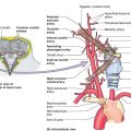
d. Thoracic duct drains the lower extremities, left trunk, left upper extremity.
e. Right lymphatic duct drains the right head/neck region, right upper extremity, right thorax.
B. Function
1. Transport interstitial fluid into the vascular system.
2. Transport chylomicrons, triglycerides, and fat from the GI tract into the blood stream through the thoracic duct.
3. Clears foreign material and particles to present them to the immune system.
II. LYMPHEDEMA
A. Definition: Accumulation of protein-rich fluid within the interstitial space due to a decrease in at least 80% of lymphatic function.
1. Postmastectomy Lymphedema:
a. Up to 10% women after breast cancer surgery have lymphedema.
b. More prevalent in cases involving XRT or radical mastectomies.
c. Thought to be less prevalent due to increased used of sentinal lymph node biopsy.
d. Presentation is a swollen, painful arm, which is at increased risk for infection.
2. Chronic lymphedema: Caused by persistent inflammation and fibrosis which then cause fatty and fibrous tissue deposition.
3. Common locations
a. Lower extremities (90%)
b. Upper extremities (10%)
i. Occurs in at least 8% of women after mastectomy
ii. Occurs in at least 35% of women after axillary node dissection and radiation
c. Genitalia (<1%)
1. Venous stasis
2. Conditions of fluid overload
a. Congestive heart failure
b. Renal disease
c. Liver disease
3. Diminished intravascular protein content
C. Diagnosis
1. History and physical examination
a. Recent surgery or trauma
b. Travel to a foreign country: Concern for Wuchereria bancrofti (filariasis)
c. Malnutrition
d. Examination
i. Nonpitting edema that usually affects hands/feet
ii. Usually not painful
iii. Minimal pigment change or ulceration
iv. Often unilateral (can measure limb circumference to document progression of disease)
v. Fluid protein content is 1 to 5 g/dL
vi. Positive stemmer sign: Inability to grasp the base of the second toe or finger due to thickening of the subcutaneous tissues.
2. Diagnostic studies
a. Lymphoscintigraphy (Gold standard test): Sensitivity: 97%; specificity: 100%
b. Nuclear medicine study which maps the lymphatic system and can identify points of blockage
c. Lymphangiogram: Can cause worsening of disease due to injection of contrast.
d. CT scan: Can differentiate lymphedema from venous stasis based on thickness of skin/muscular compartment and appearance of fluid.
e. Doppler/US: Use to rule out other causes of asymmetrical limbs (e.g., deep venous thrombosis, venous stasis).
3. Classification
a. Primary
i. Congenital (15%). Milroy’s disease: Congenital maldevelopment of lymphatic system causing lymphedema and steatorrhea, defective cell-mediated immune system, familial disease.
ii. Lymphedema praecox (75%)
a) Onset at 1 to 35 years of age
b) Often occurs with puberty
c) Most often affects the lower extremity
iii. Lymphedema tarda (10%): Onset at greater than 35 years of age
b. Secondary. Caused by a mechanical obstruction/anatomic dysfunction of an otherwise normal lymphatic system
i) Infection: Wuchereria bancrofti
ii) Iatrogenic
a) Trauma to lymphatic channels
b) Excision of lymph nodes
c) Stenosis of channels due to radiation
iii) Trauma
iv) Other: Autoimmune diseases, malignancy (primary or metastasis to lymph nodes), benign growths
4. Complications
a. Cellulitis
i. Can progress aggressively to sepsis and death
ii. Initiate treatment immediately with oral antibiotics
iii. Have low threshold to start IV antibiotic treatment
iv. Consider prophylactic antibiotics in patients who have greater than three episodes of cellulitis per year
b. Ulceration (more common in venous stasis)
c. Thickening of skin/verrucous skin changes
d. Lymphangiosarcoma (<1% incidence): Must biopsy all concerning lesions
5. Treatment
a. Elevation with proper skin hygiene
i. Use low pH solutions and water-based products
ii. Useful for most patients
b. Compression
i. Bandages, stockings, massage, pneumatic compression devices
ii. Can reduce the volume of the extremity by 25% to 60% depending on the device used.
c. Surgical management
i. Indications
a) Significant loss of function
b) Frequent infections
c) Failure of conservative/medical management
d) Significant psychological morbidity
ii. Surgical options
a) To restore lymphatic flow
1) Lymphangioplasty
2) Flap transposition
3) Microsurgical anastomosis ± microlymphatic grafting
• Anastomosis of lymphatics to veins is most common technique; lymph node transfer is gaining in popularity
• Traditionally considered not reliable; gaining renewed interest of late
b) To excise excess tissue
1) Charles operation: Excision of all tissue to the level of the muscle with skin grafting for coverage; uncommonly performed today.
2) Staged subcutaneous excision: Skin flaps are maintained, only subcutaneous tissue is excised, most commonly performed excisional technique.
3) Suction-assisted lipectomy: Liposuction techniques are used to remove subcutaneous tissues, commonly in the upper extremity and in early disease stages.
TATTOOS
I. DEFINITION: Foreign material injected into the skin which penetrates the dermis and leads to a visible/permanent mark on the skin
II. USE OF TATTOOS
A. Decorative tattoos
1. Professional—explosive growth in prevalence in the United States in last generation (one-third of all adults under age 30 have at least one); carbon base used for black dyes, variety of metals for other color pigments.
2. Amateur—variable depth; wider range of particle sizes, with larger particles refractory to laser
B. Cosmetic tattoos: Used to accentuate eyelids/eyebrows, mimic hair. Ferrous oxide is the pigment commonly used.
C. Reconstructive tattoos
1. Nipple-areolar complex reconstruction: Perform 2 to 3 months after implant-based reconstruction/flap reconstruction is complete.
2. Vitiligo: Especially useful if large, focal areas are involved
III. METHODS OF TATTOO REMOVAL
A. Destruction of tattoo: Removal of epidermis and superficial dermis
1. Nd:YAG laser
2. Infrared coagulator
4. Dermabrasion
5. Cryosurgery
6. Dermaplaning
B. Inflammatory removal: Induces removal or diffusion of pigment
1. Tannic acid
2. Oxalic acid
3. Urea
C. Surgical excision: Necessitates surgical scar in exchange for removal of the tattoo
1. Simple excision with primary closure for small tattoos
2. Excision with flap/graft coverage for larger ones
D. Laser removal of particular tattoo color pigments:
1. Red, orange, and yellow: Q-switched Nd:YAG (532-nm) or pulsed dye (510-nm) lasers
2. Black, blue, and green: Q-switched alexandrite (755-nm) and Q-switched ruby (694-nm) lasers.
3. Black: Q-switched Nd:YAG (1064-nm) is optimal for black pigment.
PEARLS
1. If the lymphedema progresses, application of intermittent pneumatic massage unless the extremity is infected, in which case massage is deferred until the infection resolves. Manual lymph compression enhances drainage through unaffected lymphatics.
2. The lymphatic system is responsible for transporting protein and lipids from the inter-stitial space to the vascular system.
3. Q-switched Nd:YAG and Alexandrite lasers helpful for tatoo removal.
4. Pulsed dye laser (585 nm, 450 ms): treatment of choice for most vascular lesions.
QUESTIONS YOU WILL BE ASKED
1. What is the difference between timing of presentation of hemangiomas versus vascular malformations?
Vascular malformations are present at birth and continue to grow as the child ages. They do not involute. Infantile hemangiomas present after birth and continue to grow until approximately one year of age after which time they often begin to involute. The exception is congenital hemangiomas which are present at birth.
2. By what age do most hemangiomas involute?
Most hemangiomas involute by 5 to 7 years of age. Approximately 50% of lesions involute by age 5. Approximately 70% of lesions involute by age 7.
3. What are the indications to perform operative intervention on a hemangioma?
1. Obstruction or deformation of critical structures such as the eye or airway.
2. Bleeding or ulceration that is unresponsive to pharmacologic management. 3. Easily excisable area with potential for an acceptable scar.
4. What nerve distribution do port-wine stains usually affect?
V1 and V2 branches of cranial nerve V.
5. Name the different syndromes associated with hemangiomas and vascular malformations along with associated signs.
See Sections IIF, IIIC, and IIIF for details.
Recommended Readings
Chim H, Drolet B, Duffy K, Koshima I, Gosain AK. Vascular anomalies and lymphedema. Plast Reconstr Surg. 2010;126(2):55e-69e. PMID: 20679788
Pohl L, Kaiser K, Raulin C. Pitfalls and recommendations in cases of laser removal of decorative tattoos with pigmented lesions: case report and review of the literature. JAMA Dermatol. 2013;149(9): 1087-1089. PMID: 23903803
< div class='tao-gold-member'>