“Silicone breast implants are the most widely used medical devices for breast reconstruction and augmentation, and revision, but even after more than 60 years of use they are associated with multiple continued complications. Using advancement in current technologies, researchers are attempting to create an optimal implant surface for patients. Through these efforts, plastic surgeons and material researchers have made great progress in the field of implant research. Multimodal techniques for the functional modification of implant surfaces will contribute to further the development of ideal biomaterials useful in breast implants.
Key points
- •
Based on the valuable efforts of plastic surgeons and material researchers, the field of implant research has made great progress.
- •
Due to advances in technologies, various attempts to create an optimal breast implant surface for patients are progressing.
- •
Multimodal techniques for the functional modification of the properties of these implant surfaces will contribute to the establishment of a variety of device choices for patients, significantly reduce complications associated with implants, and lead to the production of implants as ideal biomaterials.
Introduction
Silicone breast implants are the most widely used medical implants for breast reconstruction and augmentation, but may be associated with complications, such as Breast Implant-Associated Anaplastic Large Cell Lymphoma (BIA-ALCL), capsular contracture among others. However, the demand for silicone breast implants remains high.
Problematic materials such as semi-solid and solid materials including polyurethane, polytetrafluoroethylene (Teflon), polyvinyl alcohol formaldehyde, paraffin, petroleum jelly, and liquid silicone have been previously used as implant materials. , Although inflatable saline-filled implants, which can be placed via small incisions, are available, they do not replicate the softer, more viscous feeling of natural breast tissue. The first thick smooth silicone elastomer used was a 2-piece envelope with a seam margin. Using this initial smooth-type implant surface, an anatomically shaped implant was prepared by imitating the natural contour of the upper pole slope and curved lower pole, which is the natural female breast profile.
Because these smooth-type implants have a limited ability to maintain their proper position in vivo, Dacron patches were introduced on the posterior side of the implants to help hold their position in the pocket. However, these initial smooth implants exhibited a high capsular contracture rate and rupture at the patch interface, prompting implant manufacturers to develop the next generation of implants. Later, textured-type implants were used to reduce implant capsular contracture; however, these materials induced other issues such as late seroma, double capsule formation, and BIA-ALCL. , Thus, continuous changes in implant technology have inevitably been associated with continued limitations. ,
Modern silicone-based breast implants are composed of 2 components: silicone gel as the filling material and a silicone elastomer shell. The silicone gel functions as a filler with various viscosities depending on the length and degree of cross-linking of their polymer chain, and the silicone elastomer shell can be produced to have various roughness textures depending on the size of the pore diameter and depth. Based on advances in manufacturing technologies, various attempts to create an optimal implant surface for patients are ongoing.
The study of various properties of implant surfaces
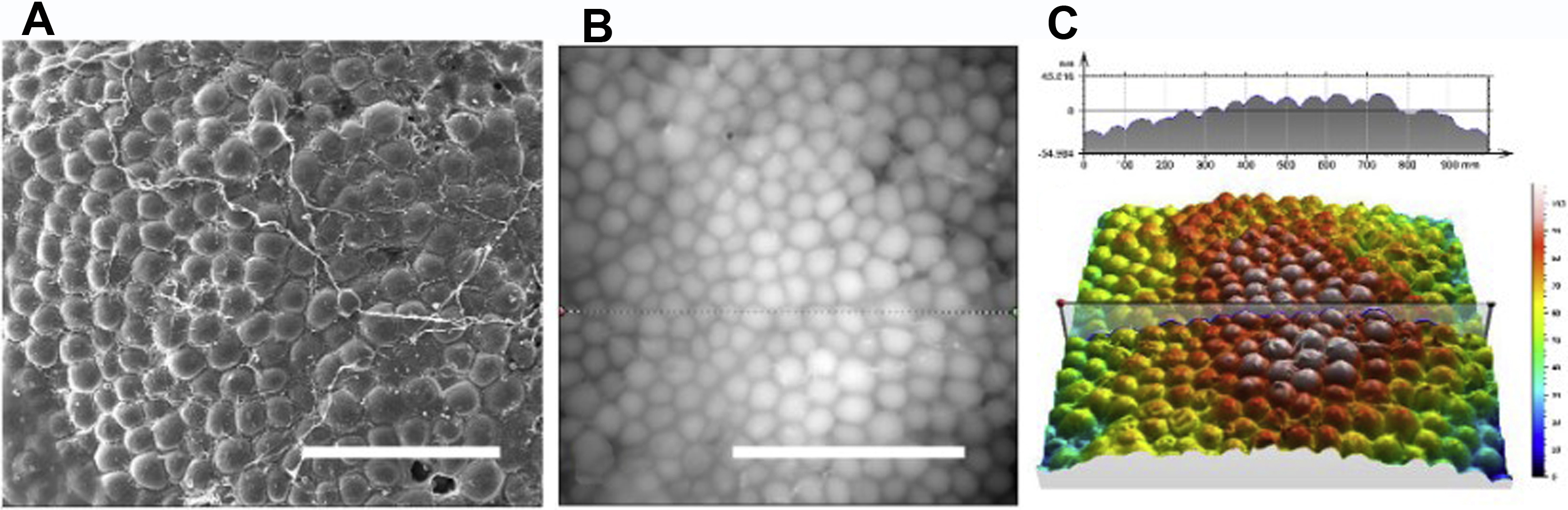
There were several classification systems for breast implant types. The most widely used classification is the International Organization for Standardization (ISO) 14607:2018. According to ISO classification, surface roughness value below 10 μm was classified as smooth type, 10 μm to 50 μm classified as microtextured type, and above 50 μm was classified as macrotextured type. A number of recent studies have presented surface classification by roughness, surface area, wettability, macrophage polarization, fibroblast activity, and bacterial adherence propensity. In addition to this, pore density, pore opening area, and depth were quantified and grouped based on surface topographic appearance and depth using scanning electron microscope images (electron beam accelerating voltage of 5 kV) and X-ray CT. The ultimate goal of these various classifications is to predict and control various complications arising from implants.
Various physical surface implants have been developed and reported to overcome implant-related complications. Some studies suggested that implants with small and fine nanotextured surfaces induce fewer complications such as seroma, infection, hematoma, dehiscence, rupture, and malposition compared with conventional implants. , These implant surfaces are classified as “smooth” according to ISO 14607:2018 classification system. The nanotextured surface consists of smaller and finer pores and “roughness” than that of other implants, increasing the number of contact points with the tissue and may be responsible for reducing dislocation and contracture. The in vitro characteristics of the implants presented in this study showed much lower roughness and height on the nanotextured surface compared with those on the microtextured surface that consist of a much larger number of contact points per unit area.
This nano/micro scale surface texturing produces significant and clear changes in cell responses. Controlling cell functions on the nano/micro scale substrate is an important focal point of materials science research. Fibroblasts, endothelial cells, and smooth muscle cells showed a clear reaction when the polydimethylsiloxane substrate was changed from the micrometer to the nanometer scale. More filopodia were observed in the nanometer groove than in the micrometer groove surface. These responses were also detected in Schwann cell, and T-cells involved in the potential capsular contracture and BIA-ALCL, which may contribute to these potential complications caused by breast implants.
Topographic modification of the silicone surface markedly affects cellular responses as well, and a more biocompatible material surface can be prepared to control these responses. However, it is important to evaluate whether the reaction of the cell at the surface leads to a more favorable tissue formation to identify a suitable method to demonstrate this relationship.
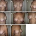
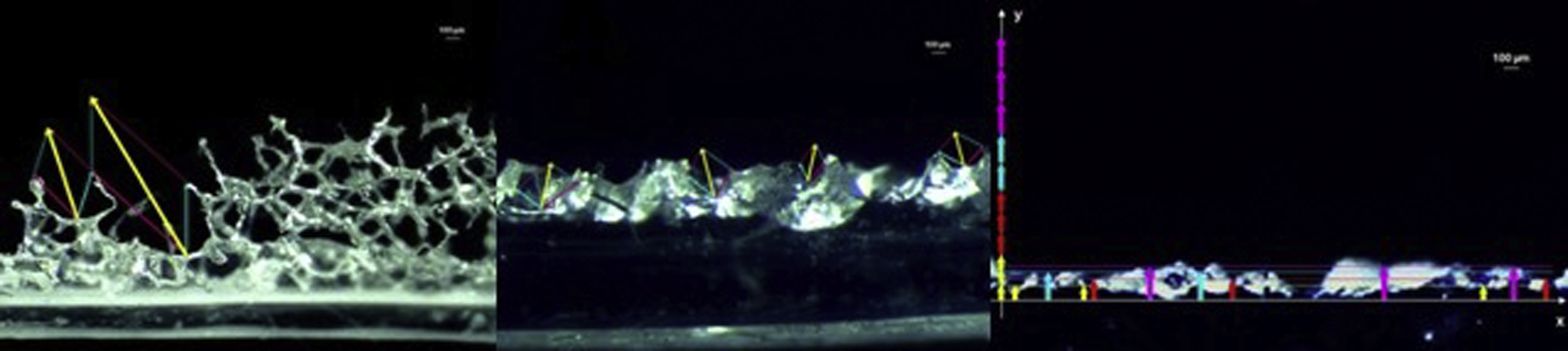
A previous study was conducted to analyze the tissue vector in the capsule tissue that formed 18 months after the insertion of implants with various pore diameters and depths. The parallelogram law presented in this study shows that vectors of various directions and lengths in capsule tissue formed by macrotextured-type implants can reduce capsular contracture ( Fig. 1 ). Based on these results, texturing below a certain level may not induce the tissue formation or integration desired. A study performed 30 years ago demonstrated that surface texturing below a certain level does not affect tissue formation, which can form various vectors.
It remains difficult to determine whether the current nano/microtextured surface is superior and represents a true advancement compared with the smooth surface. Although much data has accumulated over the years, this physical surface requires further analysis.
Recent multimodal techniques for functional modification of silicone implants
In general, most research has focused on implant silicone fill characteristics. There is a shift now based on these new studies, where functional surfaces have been developed and novel implant surfaces are emerging. Surface research is clearly a newly developing field compared with previous studies of implant size, shape (round vs anatomic), gel characteristics, filling ratio, texturing (smooth vs macro/micro/nanotextured), and roughness. Previous implant studies tended to focus on the surface texture, simple systemic or topical administration of various drugs that reduce fibrosis and inflammation, materials such as the acellular dermal matrix, and fat grafts. Our current studies have focused more on the functionalized modification of the implant surface.
Biomimetic topographic modification
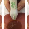
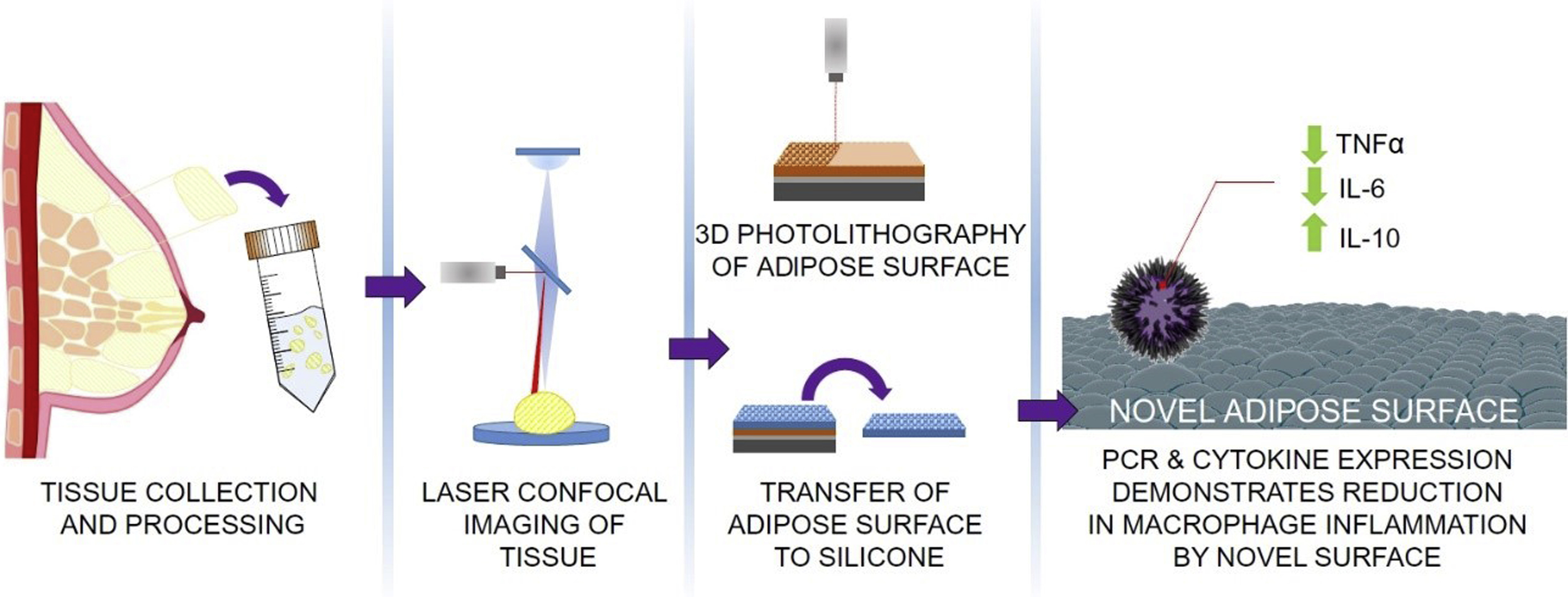
Methods for applying the topography of human tissues to implant surfaces have been developed from the previous level of simply producing texturing with nano/microscale. By analyzing topography of breast adipose tissue, a surface similar to human tissue was predicted to induce a more favorable response in the host ( Figs. 2 and 3 ). At the cellular level, results have shown that the levels of proinflammatory genes (interleukin [IL]-β1, tumor necrosis factor-α, and IL-6) were reduced and those of anti-inflammatory genes (IL-10) were increased. Fibroblasts showed a spindle-shape and spreading pattern, with a favorable response to macrophages.
Another study performed the developmental and functional evaluation of biomimetic silicone surface modification, along with the replication of the micro/nano-topographical features of the acellular dermal matrix. The dermis of the patient was de-cellularized and used in the study. The goal of the study was to produce a more human tissuelike surface rather than a simple smooth- or textured-type implant surface. Morphologic modification of the silicone surface clearly influenced the cell response, and further development of more biocompatible material surfaces may be achieved by controlling these features and extending the cell response for favorable tissue formation.
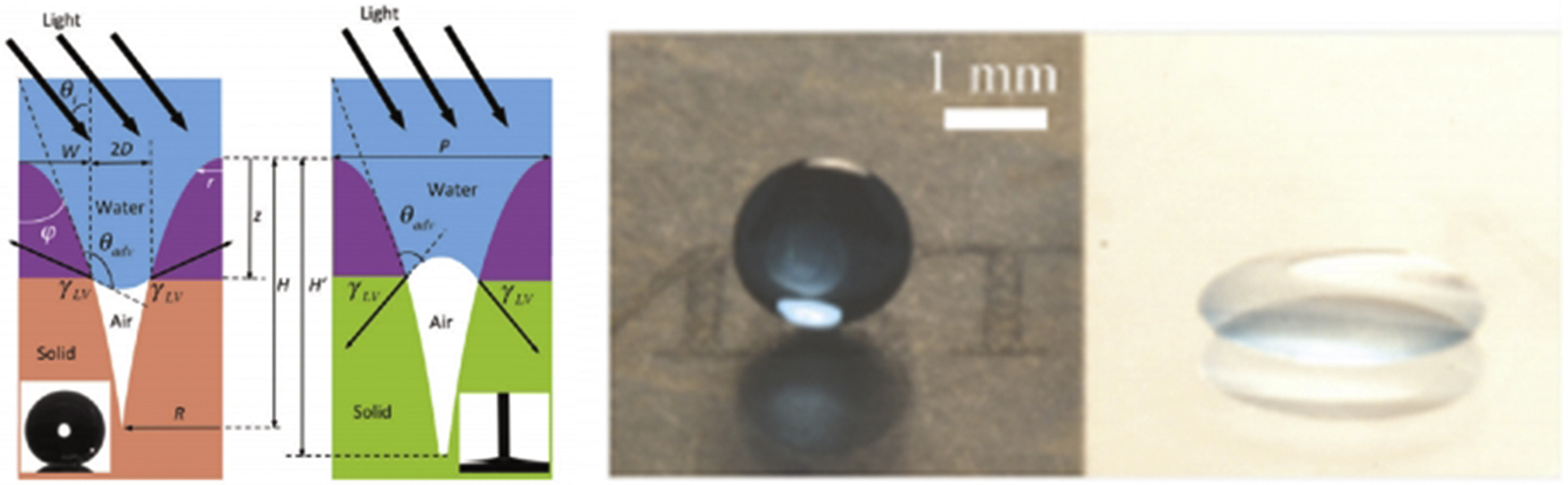
Modification of hydrophobic surface
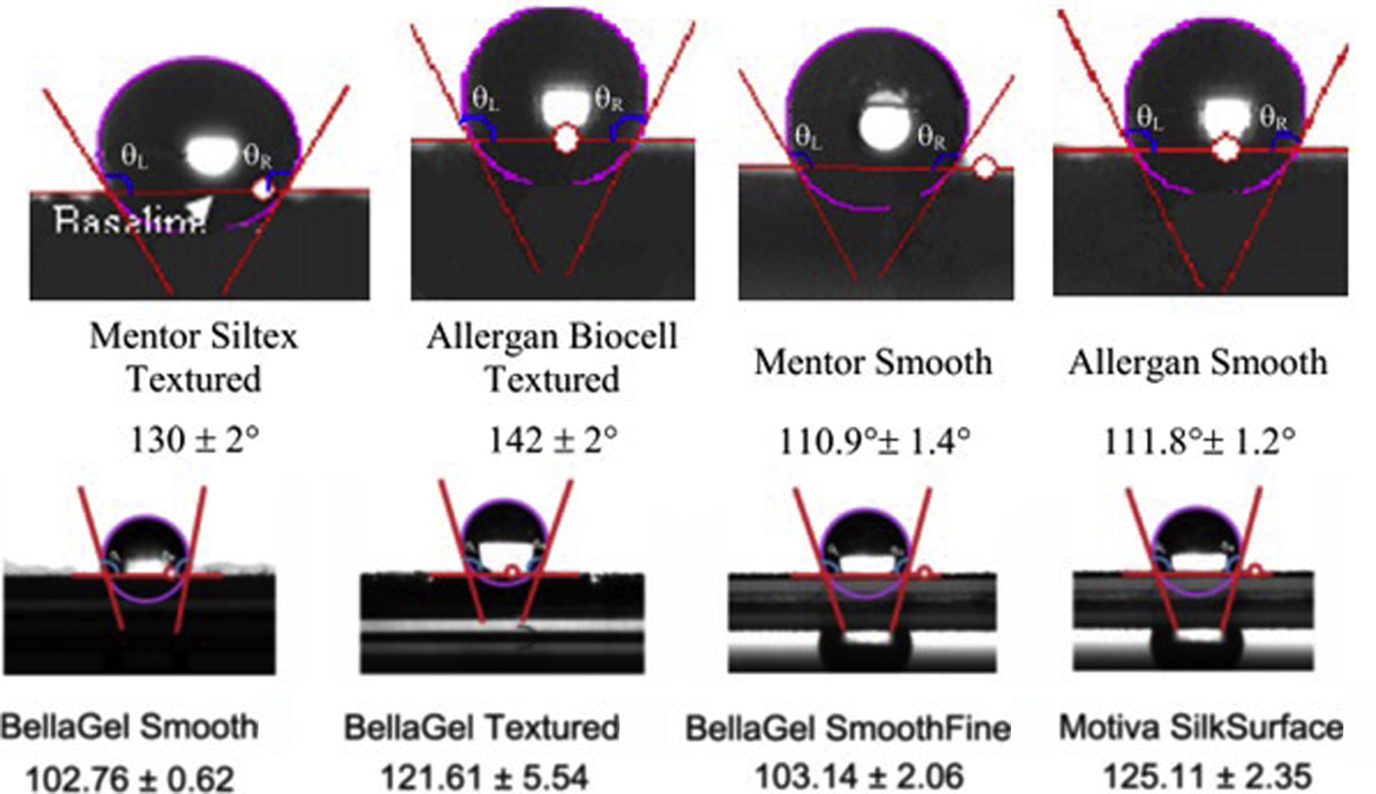
The surface contact angle is a very important indicator of surface characteristics. The implant surface has various characteristics, but most studies have been limited to its physical features. Objective data can be used to determine whether the material surface is hydrophilic or hydrophobic. When the surface of the material is hydrophobic, water that comes into contact with the surface forms rounded drops and the contact angle increases to become obtuse ( Fig. 4 ). Silicone itself is hydrophobic in nature. The contact angles of most types of implant surfaces on the market are approximately 100 to 130 , ( Fig. 5 ). Numerous studies have shown that hydrophobicity on the surface of biomaterials increases macrophage and T-cell–related inflammatory and foreign body responses , and results in the increase in the abundance of foreign body granular cells, cell fusion, and cytoskeleton formation.