7 Urodynamics: Cystometry and Urethral Function Tests
The term urodynamics means observation of the changing function of the lower urinary tract over time. Two principal methods of urodynamic investigation are known. Conventional urodynamic studies take place in the urodynamic laboratory and involve filling the bladder via a catheter, usually at a specified rate. Ambulatory urodynamic (AUD) studies are defined as a functional test of the lower urinary tract, using natural filling and thus reproducing the subject’s everyday activities. Urodynamic tests have been slow to achieve acceptance and are by no means universally used; however, in recent years there has been a resurgence of interest in the hydrodynamic and neurophysiologic aspects of the storage and evacuation of urine. An abundance of new diagnostic procedures, methodologies, and testing equipment has made it exceedingly difficult for the clinician to decide what tests are necessary to adequately evaluate lower urinary tract dysfunction in women.
To understand the fundamental value of urodynamics, one should realize that the female bladder responds similarly to various pathologies. Symptoms do not always reflect accurately the physiologic state of the bladder. For example, a patient may feel that her bladder is full when it is nearly empty, or that her bladder is contracting when it is not. Nevertheless, the evaluation of a woman with lower urinary tract complaints should not exclude the basic history and physical examination. The validity of any urodynamic diagnosis is linked to the patient’s symptoms and the reproduction of these symptoms during the testing session. To obtain the most accurate, clinically relevant interpretation of urodynamic studies, the urodynamicist should clearly understand lower urinary tract function and correlate urodynamic data with other clinical information. Ideally, the urodynamicist should be the physician who takes the history, performs the physical examination, interprets other tests, explains the diagnosis, and develops a reasonable management plan.
Results of urodynamic investigations should be recorded in a way that can be communicated among physicians and other health care personnel. For this reason, the recommendations detailed in the standardization reports of the International Continence Society (ICS) should be followed (see Appendices A and B).
Chapters 7 and 8 discuss urodynamic modalities used in the evaluation of filling, storage, and evacuation of urine. The intent is to give the reader a clear understanding of the rationale, technique, use, and limitations of each test.
PRINCIPLES OF CYSTOMETRY
The first cystometer dates back to 1872 when Schatz accidentally discovered a crude technique for measuring bladder pressure while trying to record intra-abdominal pressure. Shortly thereafter, in 1876, DuBois studied the effects of changes in body position on intravesical and intrarectal pressures and observed that the desire to void was associated with contraction of the detrusor muscle. The currently popular water cystometer was designed by Lewis in 1939. The later use of air and carbon dioxide as filling media further simplified the procedure.
Cystometry is a urodynamic test that measures the pressure and volume relationship of the bladder and is used to assess detrusor activity, sensation, capacity, and compliance. Every factor has unique implications, and before any definitive conclusions can be reached, each parameter must be examined in association with symptoms and clinical findings.
A normal bladder has the power of accommodation; it can maintain an almost constant low intravesical pressure throughout filling, regardless of volume. A normal woman should be able to suppress voiding even at maximum capacity. Then, in an acceptable environment, she should be able to initiate a voiding reflex that involves a detrusor contraction and a complete relaxation of the urethra and pelvic floor musculature.
The basic principle of cystometry is the coupling of a manometer to the bladder lumen. A filling medium is instilled into the bladder and, as it fills, intravesical pressure is measured against volume. Testing apparatuses range from simple single-channel methods, which are performed manually or electronically, to complex methods combining electronic measurements of bladder, abdominal, and urethral pressure, together with electromyography (EMG) and fluoroscopy.
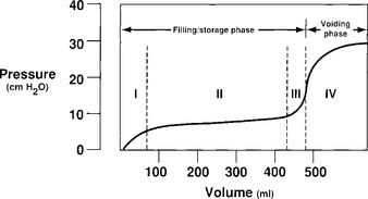
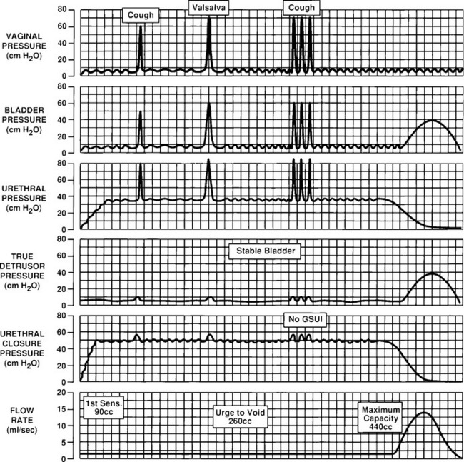
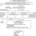
A cystometrogram has two phases: a filling/storage phase and an emptying (voiding) phase (Fig. 7-1). The filling phase is subdivided into a brief initial rise in pressure to achieve resting bladder pressure, followed by a tonus limb that reflects viscoelastic properties of accommodation of the smooth muscle and collagen of the bladder wall. There may be a third increase in the pressure, which is attributed to the stretching of detrusor muscle and collagenous elements of the bladder wall beyond their limits at bladder capacity. During this third stage, the patient is still able to suppress voiding. A detrusor contraction is then initiated voluntarily in conjunction with a relaxation of the outlet, and the patient voids.

Figure 7-1 Normal cystometrogram. Note filling phase is divided into an initial slight rise in bladder pressure (phase I), followed by a tonus limb reflecting bladder accommodation (phase II). At maximum capacity, the detrusor muscle and elastic bladder wall tissue are stretched to their limits, causing a rise in bladder pressure (phase III). A detrusor contraction is then initiated voluntarily and the patient voids (phase IV).
(Modified with permission from Wein AJ, Barrett DM. Voiding Function and Dysfunction. Mosby, Chicago, 1988.)
STANDARDIZED TERMINOLOGY AND NORMAL CYSTOMETRIC PARAMETERS
The ICS has recently redefined certain terms that are used in the reporting of cystometric results.
Intravesical pressure is the pressure within the bladder. Abdominal pressure is taken to be the pressure surrounding the bladder; it is generally estimated from rectal, vaginal, or, less commonly, extraperitoneal pressure or a bowel stoma.
Detrusor pressure is the component of intravesical pressure that is created by both active and passive forces in the bladder wall; it is estimated by subtracting abdominal pressure from intravesical pressure.
The filling phase starts when filling commences and ends when the patient and the urodynamicist decide that “permission to void” has been given. The rate at which the bladder is filled is divided into either a physiologic filling rate or a nonphysiologic filling rate. A physiologic filling rate is defined as a filling rate less than the predicted maximum filling rate—that is, the body weight in kilograms divided by 4 and expressed in milliliters per millimeter. Bladder storage function should be described according to bladder sensation, detrusor activity, bladder compliance, and bladder capacity. First sensation of bladder filling is when a patient becomes aware of the bladder filling. First desire to void is when a patient would pass urine at the next convenient moment. Strong desire to void is a persistent desire to void without the fear of leakage. Increased bladder sensation is defined as an early, first sensation or an early, strong desire to void, which occurs at low bladder volumes and which persists. Reduced bladder sensation is diminished sensation throughout bladder filling. Absent bladder sensation means that during filling cystometry, the individual has no bladder sensation. Nonspecific bladder sensations may make the individual aware of bladder filling, for example, abdominal fullness or vegetative symptoms. Bladder pain is a self-explanatory term and is an abnormal finding. Urgency is a sudden, compelling desire to void. The ICS no longer recommends the terms motor urgency and sensory urgency. These terms are often misused and have little intuitive meaning.
Compliance (C) is the change in volume for a change in pressure; it is calculated by dividing the volume change (ΔV) by the change in detrusor pressure (ΔPdet) during that change in bladder volume (C = ΔV/ΔPdet). Compliance is expressed in milliliters per centimeter.
Cystometric capacity is the bladder volume at the end of the filling cystometrogram when permission to void is usually given. The cystometric capacity is the volume voided together with any residual urine. Maximum cystometric capacity is the volume at which the patient feels she can no longer delay micturition. Maximum anesthetic bladder capacity is the volume to which the bladder can be filled under deep general or spinal anesthesia. The speed of filling, filling time, filling pressure, and type of anesthesia should be specified.
A female bladder normally experiences a first desire to void at a volume of approximately 150 to 250 mL, a normal desire to void at 300 to 400 mL, and a strong desire to void at 400 to 600 mL. During filling, an initial rise in true detrusor pressure between 2 and 8 cm H2O usually occurs. The average pressure rise is approximately 6 cm H2O and normally never exceeds 15 cm H2O. Provocation of a normal bladder by rapid filling, change of posture, coughing, or catheter movement should not incite any abnormal rises in detrusor pressure.
The urethral closure mechanism during storage may be competent or incompetent. Normal urethral closure mechanism maintains a positive urethral closure pressure during bladder filling even in the presence of increased abdominal pressure, although it may become overcome by detrusor overactivity. Incompetent urethral closure mechanism allows leakage of urine in the absence of a detrusor contraction. Urethral relaxation incontinence is leakage caused by urethral relaxation in the presence of raised abdominal pressure or detrusor overactivity. Urodynamic stress incontinence is noted during filling cystometry and is defined as the involuntary leakage of urine during increased abdominal pressure in the absence of a detrusor contraction.
EQUIPMENT
A discussion of all commercially available cystometers is beyond the scope of this chapter, but reviews by Blaivas (1990) and Rowan et al. (1987) provide an overview of available urodynamic machines. The simplest cystometer is a water manometer connected by a Y tube to both a reservoir and a catheter. A variation of this technique is discussed in Chapter 5.
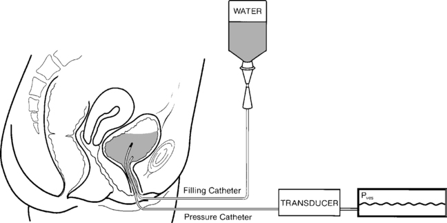
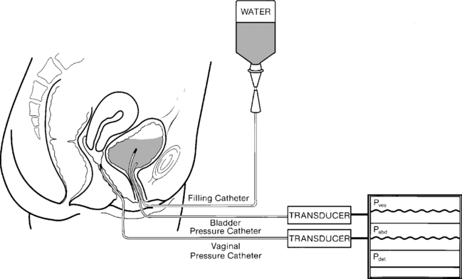
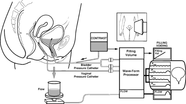
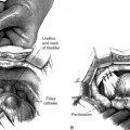
Commercially available cystometers can be broadly classified into single-channel and multi-channel machines (subtracted cystometry). Single-channel cystometry involves the placement into the bladder of a pressure-measuring catheter that produces an electronic signal, creating a graph on a recording device (Fig. 7-2). Multichannel cystometry relies on the measurement of both abdominal (Pabd) and intravesical pressures (Pves), thereby enabling one to distinguish changes in intra-abdominal pressure from changes in intravesical pressure (Fig. 7-3). Abdominal pressure can be measured via either transrectal or transvaginal catheters or less commonly from extraperitoneal pressure or a bowel stoma. We prefer vaginally placed catheters because they are more comfortable and easier to clean and maintain, and measurements are not cluttered by rectal peristalsis. Electronic subtraction of intra-abdominal from intravesical pressure allows for the calculation of true detrusor pressure (Pdet).

Figure 7-3 Subtracted cystometry. Intravesical and intra-abdominal pressures are measured, and true detrusor pressure is electronically derived (Pves – Pabd). Pves, Bladder pressure; Pabd, abdominal pressure; Pdet, detrusor pressure.
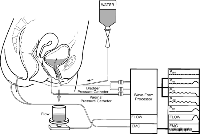
Subtracted cystometry may be enhanced further by additional measurement of urethral pressure (Pure). This measurement allows for the calculation of urethral closure pressure (Pucp), which is the difference between urethral and bladder pressures. Certain machines also allow for the simultaneous measurement of EMG activity and the performance of flow studies (Fig. 7-4). These standard urodynamic techniques may be combined with videocystourethrography, which is termed video urodynamics.

Figure 7-4 Multichannel urodynamics. Intravesical, intra-abdominal, and intraurethral pressures are measured. True detrusor pressure (Pdet) and urethral closure pressure (Pucp) are electronically derived. EMG and flow studies are also performed. Pves, Bladder pressure; Pabd, abdominal pressure; Pdet, detrusor pressure; Pure, urethral pressure; Pucp, urethral closure pressure; EMG, electromyography.
METHODOLOGY
Despite the widespread use of filling cystometry, the optimal technique for performing the test is unknown. The following section addresses the various technical aspects, controversies, and techniques for performing filling cystometry.
Filling Media
The commonly used infusants for cystometry include water, carbon dioxide, and radiographic contrast material. In 1971, Merrill et al. introduced the use of carbon dioxide (CO2). Although once popular in North America, it is rarely, if ever, used for the following reasons: (1) It further decreases the physiologic nature of the test; (2) if gas is used, the bladder volume cannot be assessed because CO2 is compressible; (3) CO2 dissolves in urine to form carbonic acid, which irritates and reduces cystometric bladder capacity; (4) abdominal pressure is not usually measured during CO2 cystometry, making interpretation more difficult; and (5) when CO2 is used for filling cystometry, it is impossible to perform a stress test or voiding studies.
Water or physiologic saline is the most commonly used filling medium unless radiologic screening is also being performed, in which case contrast medium is used. The cystometric findings are not affected by the choice of liquid medium.
Position of Patient and Provoking Maneuvers
Cystometry should mimic everyday stresses on the bladder as much as possible. Thus, performing the test with the patient in the sitting or standing position is preferable. During cystometry, the bladder should be provoked by a series of tests that usually include coughing, heel bouncing, walking in place, and listening to running water. These maneuvers may provoke uninhibited detrusor contractions or induce stress incontinence.
Physical factors may influence the positioning of patients during urodynamic tests; for example, in elderly patients or those with neurologic disease, it may be difficult to undertake cystometry in any position other than supine.
Temperature of Fluid
Most laboratories use fluid at room temperature, although some investigators believe that the installation of warm or cold fluid may provoke abnormal bladder activity. The installation of ice water (Bor’s test) is occasionally used as a test for neurologic disorders.
Technique of Bladder Filling
Theoretically, the most physiologic method of filling is by diuresis, combined with a suprapubically placed pressure line. The long time needed to investigate the patient prohibits natural filling as a practical method of performing cystometry in most centers. Therefore, cystometry is usually performed through a transurethrally placed catheter. Filling is accomplished using either simple gravity or a water pump. The bladder is filled through either a small catheter or, preferably, a separate channel on the pressure-measuring catheter.
Rate of Bladder Filling
The ICS no longer divides filling rates into slow, medium, and fast. In practice almost all investigations are performed using medium filling rates that have a wide range. It may be more important during investigations to consider whether or not the filling rate used during conventional urodynamic studies can be considered physiologic.
Types of Catheters
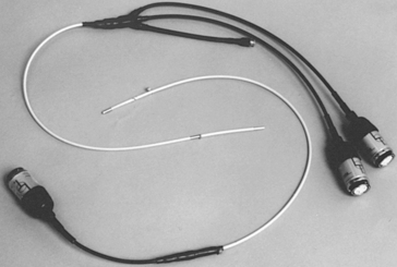
Various catheters have been used for cystometry. Simple or manual cystometry can be performed with a transurethral Foley catheter. Electronically monitored studies require more sophisticated balloon or microtransducer catheters. Water-filled balloon catheters or water perfusion catheters have been used with moderate success. These catheters are inexpensive, disposable, and easy to use. However, more sophisticated laboratories usually use sensitive microtransducer catheters (Fig. 7-5). These catheters are available with one to six microtransducers on the catheter. They have small diameters, are flexible, and can measure accurately rapid changes in pressure during repetitive coughing or other provoking maneuvers. Disadvantages include their expense, their need to be replaced after approximately 100 studies, and their tendency to produce rotational artifacts; pressure readings may vary depending on the orientation of the transducer to the bladder or urethral wall.

Figure 7-5 Two microtransducer catheters. One catheter has a single microtransducer and is used for estimating abdominal pressure. The other catheter has two microtransducers approximately 6 cm apart used to measure intravesical and intraurethral pressure. This catheter also contains a fluid-filling port.
Technique of Cystometry
The technique of subtracted urethrocystometry used in our laboratory is as follows:

Figure 7-6 Normal filling and voiding subtracted cystometry. Note that provocation in the form of coughing and straining does not provoke any abnormal rise in true detrusor pressure. At maximum capacity on command, a detrusor contraction is generated and voiding is initiated. USUI, Urodynamic stress urinary incontinence.
INDICATIONS FOR CYSTOMETRY
The indications for cystometry are somewhat controversial. Each patient must be evaluated individually. Based on clinical findings and planned treatments, the physician must decide whether cystometry is indicated and, if it is, whether it should be performed via a simple office test or with more sophisticated electronic testing. In our opinion, an electronic single-channel study does not offer any more information than does a carefully performed nonelectronic test. We believe that the only reason to perform electronic urodynamic testing is to measure pressures from several anatomic sites, thus obtaining subtracted pressures of importance.
Indications for single-channel cystometry versus subtracted or multichannel cystometry have been debated extensively; however, few comparisons exist in the literature. One study by Ouslander et al. (1987) reported a sensitivity of 75% in geriatric patients undergoing simple supine cystometry when compared with multichannel testing. Sutherst and Brown (1984) compared single-channel and multichannel urodynamics in a blinded crossover study of 100 incontinent women. They noted single-channel studies to be 100% sensitive and 89% specific compared with multichannel studies. Multichannel cystometry may have a higher sensitivity for recognizing low-pressure detrusor contractions. Multichannel techniques also improve the specificity of cystometry by avoiding false-positive test results created by increases in abdominal pressure. Whether the cost of multichannel testing is justified for most patients remains to be proved. Box 7-1 lists suggested indications for subtracted cystometry.
BOX 7-1 INDICATIONS FOR MULTICHANNEL SUBTRACTED CYSTOMETRY
Inconclusive single-channel studies
Stress incontinence before surgical correction
Urge incontinence not responsive to therapy
Recurrent urinary loss after previous surgery for stress incontinence
Frequency, urgency, and pain syndromes not responsive to therapy
Nocturnal enuresis not responsive to therapy
Lower urinary tract dysfunction after pelvic radiation or radical pelvic surgery
VIDEO–URODYNAMIC TESTING
Video-urodynamic studies of the lower urinary tract represent a combination of video-cystourethrography and standard urodynamic techniques. Video-urodynamics requires equipment for cystometry, plus an image intensifier and a videotape recorder. In addition, various interface modules are necessary, depending on the exact design of the system. A television camera positioned above the recorder with a mixing device projects the recording channels on a television monitor alongside the radiographic image of the bladder (Fig. 7-7). Radiopaque-filling medium is used for video-urodynamic studies. As with all other urodynamic studies, every effort must be made to limit the inhibitory effect of the additional machinery and personnel imposed on the patient.

Figure 7-7 Video-urodynamic testing. Multichannel urodynamic tests are performed under fluoroscopy, thus allowing simultaneous visualization of the lower urinary tract during recording of pressures. Pves, Bladder pressure; Pabd, abdominal pressure; Pdet, detrusor pressure; Pucp, urethral closure pressure.
Potential advantages of video-urodynamic studies include the consolidation of multiple evaluation modalities into one examination, thereby providing information about lower urinary tract anatomy and function under various provocative environments. Descent of the bladder neck, milk-back of urine from the urethra to the bladder, and bladder neck funneling all may be visualized during simultaneous recording and imaging of bladder, urethral, and abdominal pressures. Anatomic abnormalities, such as urethral or bladder diverticula, may also be noted. The major disadvantages of video-urodynamic testing are the radiation exposure, cost, and technical expertise and support necessary for its use.
The indications for video-urodynamic studies are controversial. Some authorities believe that no additional information is obtained when these studies are compared with more conventional nonimaged multichannel studies; however, others believe that valuable additional information can be obtained from simultaneous imaging, especially in recurrent cases of incontinence or complicated neurologic conditions. Visualizing bladder neck opening at rest and during straining may help differentiate stress incontinence secondary to bladder neck hypermobility from intrinsic sphincter deficiency.
AMBULATORY URODYNAMICS (AUD)
The largest deficiency of currently available urodynamic techniques is that laboratory observations may not always represent accurately physiologic behavior of the bladder and urethra. At times, the urodynamicist cannot reproduce the patient’s symptoms in the laboratory setting. Several companies have recently developed commercially available AUD systems (AUDS). This equipment uses indwelling catheter-mounted transducers that are connected to a microcomputer worn over the patient’s shoulder. This allows freedom of movement to the extent that the patient can reproduce the activities that incite lower urinary tract dysfunction. In principle, these systems are the same as those used for conventional urodynamics, and the same basic methodology applies. AUDS should be considered when conventional urodynamics fail to provide a pathophysiologic explanation for the patient’s symptoms. The most common example of this is in a patient who complains of incontinence that cannot be objectively demonstrated and has failed nonsurgical modes of therapy. Before a more invasive intervention, such as surgery, is considered, AUDS could be used to objectively demonstrate the incontinence. AUDS are also helpful in determining whether detrusor overactivity or urethral incompetence is the main cause of incontinence in women for whom surgery is being contemplated.
The technique for performing AUDS, as described by Abrams (1997), involves the recording of three micturition cycles: a resting cycle, when the patient sits in a chair; an ambulant cycle, when the patient moves around the hospital; and an exercising cycle, which should include any specific incontinence-provoking measures. Once the three cycles are recorded, the information is downloaded to a computer for analysis. This analysis is time-consuming and requires considerable expertise.
Besides providing objective evidence and the cause of the patient’s lower urinary tract symptoms, additional interesting information is being generated by AUDS. Present ideas on bladder compliance, detrusor overactivity, and voiding function are being questioned. AUDS studies performed on asymptomatic, neurologically intact volunteers have shown a 30% incidence of detrusor overactivity. It has been previously suspected that bladder compliance is related to the speed of bladder-filling, and AUDS has confirmed this fact (Kulseng-Hanssen and Klevmark, 1996). Finally, voiding pressures during AUDS in women have been shown to be significantly higher than those obtained during conventional urodynamics.

Stay updated, free articles. Join our Telegram channel

Full access? Get Clinical Tree

