This article offers a review of the various three-dimensional (3D) imaging devices currently on the market and further examines their distinct modalities and functionality. The authors highlight the different modalities of 3D imaging and the corresponding advantages, disadvantages, and associated costs. The authors aim with this comprehensive review is to educate providers on the utility of 3D imaging in esthetics as well as help guide them based on their specific needs and financial considerations.
Key points
- •
This article dives into the history of three-dimensional (3D) imaging and how stereophotogrammetry has been and remains the predominant modality for acquiring 3D images.
- •
A comprehensive comparison of various 3D imaging devices is presented, with each device being assessed on its advantages, disadvantages, cost, and specific modality, thereby offering invaluable insights for potential users.
- •
A review of the barriers encountered in the utilization of 3D imaging, focusing on the financial burden, technological challenges, and low availability of validation studies.
Introduction
Three-Dimensional Photography in Medical Practice
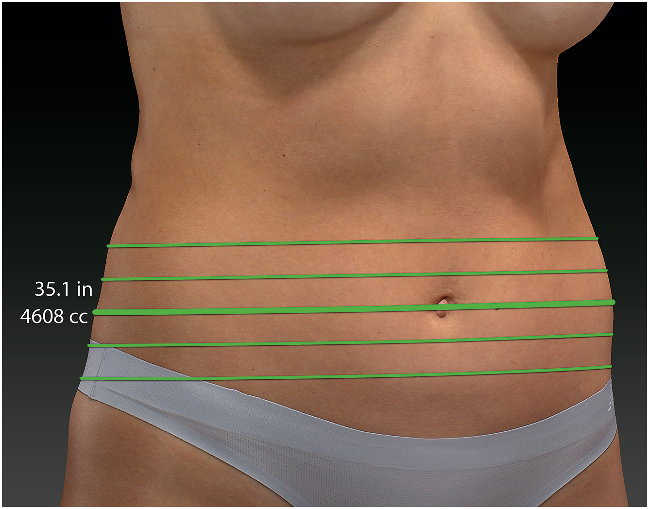
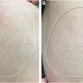
Three-dimensional (3D) photography has raised in popularity over recent years, with application in both academic and private practice settings for dermatologists and plastic surgeons. This technology has become an invaluable tool in cosmetics for presurgical patients consultation or procedural planning and postoperative assessment, offering insights into facial symmetry and tracking changes [ ]. Outside of dermatology, it has been used in orthodontics [ ], dental rehabilitation [ ], orthopedic prosthetics [ ], and both cranial–facial and maxillofacial repair [ ]. Three-dimensional photography, compared with traditional 2D photography, provides a more comprehensive visualization, incorporating surface anatomy, allowing for volumetric assessments, and offering detailed information on facial contour and shape [ , ]. It is also capable of providing topographic distance or circumferential measurements which can be beneficial in preoperative consultation ( Fig. 1 ) [ ].
Imaging Modalities
Tracing back to its initial application in 1944, 3D photography has undergone significant advancements. Initially capture 3D surface anatomy, stereophotogrammetry used measurements from 2D photographs to generate a 3D facial model through plotting machines [ ]. Presently, the technology continues to apply multiple synchronous photographs from various angles to create a detailed 3D image.
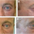
Stereophotogrammetry remains the most favored method for creating 3D photos. It complies images taken from different angles to form a cohesive 3D model. Despite the availability of other 3D modalities which are discussed later in this review, stereophotogrammetry is the preferred method due to its minimal risk profile ( Table 1 ) [ , ]. This method can use a high-resolution, rapid-acquisition camera system to create manipulatable 3D models with the capability of measuring linear and angular distances and volumetric analysis [ ]. Images can be captured by cameras wirelessly, wired, or transferred by memory card to a desktop computer/laptop which handles the heavy lifting of computation analysis, tiling and image compilation ( Fig. 2 ).
| 3D Imaging Modality | Mechanism of Action | Advantages | Disadvantages |
|---|---|---|---|
| Stereophotogrammetry | Multiple synchronous photographs at various angles to create a detailed three-dimensional image |
|
|
| Computed tomography (CT) | Uses x-rays to create cross-sectional images of the body to reconstruct 3D images |
|
|
| MRI | Uses magnetic fields to generate detailed images of the inside of the body to form 3D images |
|
|
| Ultrasound | Uses sound waves that bounce off structures in the body, creating echoes that are used to form images | Rapid User-friendly |
|
| Laser surface scanning | Uses lasers to rapidly move across the skin surface and scan to create a map of the skin |
| High financial burden |
| Structured lighting | Projects light patterns onto the surface of the item being measured. The degree of light displacement is measured and correlates to the degree of contour or elevation | 3D reconstruction through light displacement | Requires specialized equipment |
Computed tomography (CT) can create 3D photography with the ability to distinguish subcutaneous tissue from underlying muscle and fascia [ , ]. Similar to stereophotogrammetry, it can quantify volume but it exposes patients to ionizing radiation and requires supine positioning, potentially distorting the 3D image [ ].
MRI may be a safer alternative as it lacks exposure to ionizing radiation. It can be used safely in longitudinal studies requiring repeated scans [ ]. It offers precision and reproducibility but, similar to the CT scan, requires patients to lie flat. This position change can cause gravitational changes in the facial tissue [ , ]. In addition, it is limited by long wait times, inaccessibility in dermatology clinics and high costs [ ]. Because of these limitations, this method of 3D imaging is not used frequently in esthetic procedures [ ].
Ultrasound can generate images via sound wave reflection [ ]. It can quantify superficial soft tissue and has been used as an objective measure of lipoatrophy in HIV patients for clinical trials [ , ]. It can also help evaluate skin thickness. Compared with CT and MRI, it is quick, easy, and does not expose patients to ionizing radiation. However, its depth is limited and the necessary pressure from the ultrasound probe may alter the skin structure [ , ].
Laser surface scanning is another modality, providing a rapid surface view in 3D imaging. However, it cannot measure beneath the skin. This device is eye-safe and can be hand-held or mobile [ ]. Although it is easy to use, it can carry a high financial burden to have in the clinic [ ]. These lasers are thought to be the most accurate of all 3D capture systems; however, it has low clinical utility given their high cost and long capture times [ ].
Structured lighting, a method involving the projection of light patterns on the subject, offers another approach to 3D creation [ ]. This method relies on infrared sensors and specialized equipment to measure the degree of light displacement on the subject to determine the degree of contour or elevation [ ].
Review of existing three-dimensional devices
Stereophotogrammetry
Several 3D devices are currently available including prominent systems such as 3D Vectra XT, H2 (Canfield Scientific, Fairfield, NJ), Crisalix VR (Crisalix SA, Lausanne, Switzerland), 3dMD (3dMD Inc, Atlanta, GA), and FastSCAN (Polhemus, Colchester, VT) [ ]. These innovative devices allow clinicians to evaluate different parameters including symmetry and proportions, thereby crafting a visual representation of potential postoperative results ( Table 2 ).
| Device Name | Modality of 3D Imaging | Advantages | Disadvantages | Cost |
|---|---|---|---|---|
| 3D Vectra XT | Stereophotogrammetry |
|
| NR |
| Vectra H2 | Stereophotogrammetry |
|
| $13,000 |
| Vectra M3 | Stereophotogrammetry |
|
| NR |
| Crisalix VR | Structured light triangulation |
| Low utility in preoperative modeling | NR |
| 3DMD | Stereophotogrammetry | Precise and reliable | Non-portable Expensive Require structured lighting | >$25,000 |
| FastSCAN | Laser Scanning |
|
| NR |
Related posts:

Stay updated, free articles. Join our Telegram channel

Full access? Get Clinical Tree


