EMD Chemicals, Philadelphia PA
1.4.2 Principles of Molecular Biology
1.4.3 Proteomics, genomics and epigenetics
1.4.4 Application for Skin Care
1.4.5 (Future perspectives) Conclusion
* A note from Meyer R. Rosen
Before we get started on perhaps one of the most complex chapters in this book, we call attention to the fact that many readers will not be familiar with numerous terms used in the text. To this end, a glossary has been created at the end of the chapter. To facilitate understanding, note that in the text all “big” words, unfamiliar to many, have been bolded, and a concise definition is presented in the glossary.
As Editor-in-Chief of this book—I take it as my duty to urge you, the reader (no disrespect intended) to grow in the 50-year-old tradition of Harry’s Cosmeticology. The contents of this chapter are deemed so important that, taking editorial prerogative, it has been put in the first section of the book, “In the Beginning,” along with other key areas that provide the foundation for new ingredients and products that are essential for understanding and leading innovation in our industry.
This chapter introduces the basic fundamentals of the principles of molecular biology and why cosmetic industry professionals must understand them. It is our opinion, based on the developmental path being taken by ingredient suppliers and formulators, that, like learning to effectively use social media in the Internet age, if you do not at least have a basic understanding of these concepts, you will be left behind.
Consider this analogy, which is quite prevalent at the time of this writing. When young children age three or so and older, and teenagers, are walking around with their iPads and iPhones, etc., and you—the adult, the parent, the seeker of “how do I text, or use Linkedin,” etc.—are your resource for how to do IT. If you do not use that brilliant mind of yours to lead the knowledge pack rather than be a follower, then, my friends, your foreseeable contribution to the development of new cosmetic and personal care products will wither into dust and be blown away by the wind.
And Now, to the Heart of the Matter…
The Human Genome Project revolutionized scientific research in the field of molecular biology. Many of the techniques and gene interaction knowledge base currently in use are the result of the project. The cost and time to conduct such studies continues to decrease, resulting in the generation of information relevant to the formulations chemist. Molecular cell biology investigates how cells develop, operate, communicate, and control their activities. Cells communicate with body tissue, which is composed of cells. Tissues communicate with other tissues and organs throughout the body. Generally the transfer of information between cells, tissues, and organs is through proteins. We now know that genes consisting of deoxyribonucleic acid (DNA) are responsible for biological structure, maintenance, and integration of cellular function. DNA indirectly controls the synthesis of many small molecules including proteins. DNA may be considered a storage form of genetic information. Gene expression is how cells “know” to make the right proteins at the right time in the right quantities.
1.4.2 PRINCIPLES OF MOLECULAR BIOLOGY
Amino Acids and Proteins: Their Role in Cell and Tissue Communication
Proteins are the working molecules of the cell; they carry out the program of activities encoded by genes. To do this a coordinated effort of many different types of proteins is necessary (1). The three-dimensional structure of a protein enables it to regulate concentrations of metabolites, cause cell motion, and provide structural rigidity to the cell. The spatial organization of proteins is critical in understanding how they work (1).
Proteins consist of amino acids, and there are only twenty amino acids from which proteins derive their structure. The three-dimensional structure of the protein is determined by the amino acid sequence. Amino acids can be classified primarily on their solubility in water, which is influenced by the polarity of their side chains. Amino acids with polar side groups tend to be found on the cell surface and make proteins soluble in aqueous solutions. Amino acids with nonpolar side groups avoid water and aggregate to form the water-insoluble core of the protein. Water-soluble amino acids have ionized or polar side chains. At neutral pH, arginine and lysine are positively charged; aspartic acid and glutamic acid are negatively charged and exist as aspartate and glutamate. These four amino acids are the major contributors to the overall charge of a protein (1). Histidine has a pKa of 6.8, the pH of cytoplasm in the cell.
Small shifts in the pH of the cell will change the charge of histidine side chains. The activity of numerous proteins is modulated by pH through protonation of histidine side chains. Asparagine and glutamine are uncharged and have polar amide groups with extensive hydrogen-bonding capacities. Serine and threonine are uncharged and have polar hydroxyl groups facilitating hydrogen bonding with other polar molecules. Hydrophobic amino acids have aliphatic side chains that are insoluble or slightly soluble in water. These amino acids tend to pack in the interior of proteins away from the aqueous environment. Cysteine, glycine, and proline have side chains with unique properties. The cysteine side chain contains a reactive sulfhydryl that can oxidize to form a disulfide bond to a second cysteine. Disulfide bonds are commonly found in extracellular proteins where they facilitate maintaining a folded structure. Glycine, the smallest amino acid, has a single hydrogen atom as its R group. Its small size allows it to fit into tight spaces. Proline and glycine may be found at locations on a protein’s surface where the chain loops back into the protein.
The Cell
All organisms are composed of cells. A cell is a membrane-bound unit that contains DNA and cytoplasm. A plasma membrane encloses each cell, separating its contents from its surroundings. The plasma membrane contains numerous proteins that are responsible for the ability of the cell to interact with its environment. For example, receptor proteins in the membrane interact with hormones and induce changes in the cell in response to the interaction. Transport proteins help molecules and ions move across the plasma membrane either from the membrane at the cell surface to inside the cell, or from inside the cell to the cell surface. Every cell contains DNA, which is the hereditary molecule containing the genes that code for the proteins being synthesized by the cell.
Cells found in animals referred to as eukaryotic cells contain numerous compartments termed organelles and smaller sac-like structures known as vesicles. The largest organelle in a eukaryotic cell is the nucleus, which is the repository of the genetic information that directs all the activities of a living eukaryotic cell. The nucleolus is an area found in the nucleus where intensive synthesis of ribosomal RNA is occurring. A nuclear envelope is located on the surface of the nucleus. The outer membrane of the nuclear envelope is continuous with the cytoplasm’s interior membrane system called the endoplasmic reticulum. Scattered over the surface of the nuclear envelope are nuclear pores filled with proteins that act as molecular channels permitting certain molecules to pass into and out of the nucleus. Proteins moving into the nucleus may be incorporated into nuclear structures or catalyze nuclear activities; RNA and protein-RNA complexes are formed in the nucleus and exported to the cytoplasm. DNA of the eukaryotic cell is divided into linear chromosomes, which are fully extended into thread-like strands called chromatin. This arrangement allows proteins to attach to specific nucleotide sequences along the DNA. Without this protein interaction DNA could not regulate the day-to-day activities of the cell. Chromosomes are associated with packaging proteins called histones.
When a cell is ready to divide, the DNA coils up around the histones into a tight condensed structure; this initial aggregation is termed a nucleosome, and resembles a bead on a string (2). After cell division the chromosomes uncoil, permitting RNA polymerase—an enzyme that makes RNA copies of the DNA—to gain access to the DNA. RNA copies of DNA are the only way hereditary information in the DNA can be used to direct the synthesis of proteins. The eukaryotic cell is distinguished from plant cells with the presence of internal membranes in the cell. The largest internal membrane is the endoplasmic reticulum (ER). The ER is composed of a lipid bi-layer embedded with proteins. The surface is embedded with ribosomes involved in protein synthesis. Proteins synthesize on the surface of the ER (termed rough ER because the surface appears pebbly, like the surface of sandpaper). These proteins contain special amino acid sequences called signal sequences. These protein sequences travel through the ER membrane to the interior of the ER and then move to a vesicle-forming system known as the Golgi apparatus, and is eventually released to the outside of the cell.
Eukaryotic cells contain a variety of vesicles. Lysosomes are membrane-bounded digestive vesicles that contain enzymes catalyzing the breakdown of proteins. Other vesicles, known as peroxisomes, contain the enzyme catalayse, which breaks down hydrogen peroxide into water and oxygen. The breakdown of hydrogen peroxide is important because hydrogen peroxide buildup in the cell is toxic. Mitochrondria are organelles containing proteins that carry out oxidative metabolism of the cell. They have their own DNA-containing genes that produce proteins essential to the mitochrondria’s role as centers of oxidative metabolism. Most of the genes that produce the enzymes used in oxidative metabolism are located in the nucleus of the cell. The nucleus is the control center of the cell for direction of protein synthesis and cell reproduction. The nucleolus is the manufacturing site for ribosomal subunits, which are sites for rRNA synthesis, a process necessary for DNA replication. Ribosomes provide a framework for protein synthesis.
1.4.3 PROTEOMICS, GENOMICS AND EPIGENETICS
Protein Conformations and Cell Communication
Mammalian cells contain up to 10,000 different kinds of proteins (1). Proteins must be localized to the appropriate cellular membrane or aqueous compartment to function properly (Figure 1). Proteins are the working molecules of the cell; they carry out the program of activities encoded by genes. The structure of the protein gives rise to its function. The function of virtually all proteins depends on their ability to bind to other molecules, or ligands with specificity. There are numerous types of receptors on the cell surface, and each is essential for signaling between cells. Affinity refers to the strength of binding and specificity refers to the ability of a protein to bind one molecule in preference to another molecule. In some cases a particular protein on one cell binds to a receptor protein on the surface of an adjacent cell, triggering differentiation.
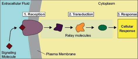
Cell signaling can be divided into three stages.
1. Reception: A cell detects a signaling molecule from the outside of the cell. A signal is detected when the chemical signal (also known as a ligand) binds to a receptor protein on the surface of the cell or inside the cell.
2. Transduction: When the signaling molecule binds the receptor, it changes the receptor protein in some way. This change initiates the process of transduction. Signal transduction is usually a pathway of several steps. Each relay molecule in the signal transduction pathway changes the next molecule in the pathway.
3. Response: Finally, the signal triggers a specific cellular response.
Source: Overview of Cell Signaling. http://creativecommons.org/ns#” xmlns:dct=“http://purl.org/dc/terms/” about=“http://www.hartnell.edu/tutorials/biology/signaltransduction.html
Available at: http://www.hartnell.edu/tutorials/biology/signaltransduction.html
Signal molecules are designated, endocrine, autocrine, or paracrine depending upon the distance over which the signal acts. Membrane-bound proteins on a neighboring cell can also signal to each other. In endocrine signaling the signaling molecules, or hormones, act on target cells distant from their site of synthesis. In paracrine signaling the molecules released are in close proximity to each other. In autocrine signaling cells respond to substances that they release themselves. There are compounds that may act by multiple types of signaling. Epidermal growth factor (EGF) is an example of a protein hormone synthesized in the plasma membrane. Membrane-bound EGF can bind to and signal an adjacent cell by direct contact. Cleavage by a protease releases secreted EGF, which then acts as an endocrine signal on distant cells (1). There are four major classes of cell-surface receptors: 1) receptors with enzymatic regulatory function; 2) G-protein-coupled receptors, which activate or inhibit an enzyme that generates a specific second messenger or modulates an ion channel, causing a change in membrane potential; 3) ion channel receptors, which change the conformation of the receptor to enable specific ions to flow through the receptor resulting in a change of the electric potential across the cell membrane; and 4) tyrosine kinase-linked receptors, in which ligand binding stimulates formation of a dimeric receptor, which interacts with and activates one or more cytosolic protein tyrosine kinases. Receptors for many cytokines, interferons, and human growth factor are of this type. (See Figure 1)
GTPase
Related posts:
 – AN OVERVIEW OF THE CHANGING REGULATORY LANDSCAPE IN THE U.S. AND THE E.U. AND HOW TO DEAL WITH THEM…
 – ACHIEVING GLOBAL MARKET ACCESS- FOCUS ON RUSSIA
 – CLASSIFICATION SCALE FOR SKIN COMPLEXIONS AROUND THE WORLD
 – ASIAN ETHNIC SKIN: SPECIALTY CORRECTIVE COSMECEUTICALS FOR ASIAN ETHNIC SKIN CARE
 – THE NOSE: ACCESSING THE BIOLOGY OF HUMAN OLFACTION: NEW, ALL-NATURAL FRAGRANCE INGREDIENTS; NOVEL CONSUMER FRAGRANCE EXPERIENCES AND APPLICATIONS
 – MECHANISMS OF CHANGES IN HAIR SHAPE
Stay updated, free articles. Join our Telegram channel

Full access? Get Clinical Tree


