FIGURE 16-1 Diagnostic ultrasound being used on medial thigh to detect vascular occlusion
TABLE 16-1 US Technology Use
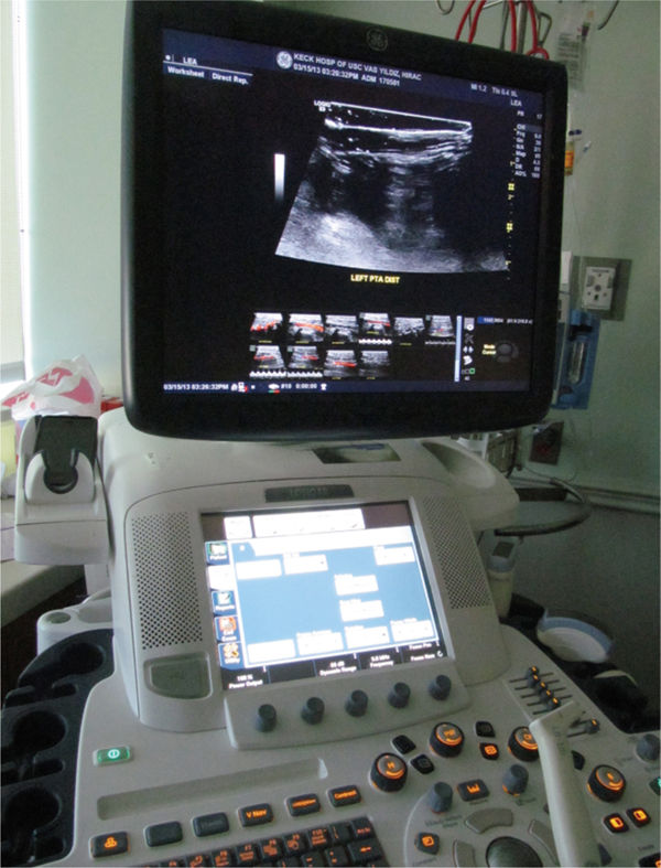
FIGURE 16-2 Diagnostic ultrasound equipment used for peripheral vascular studies Diagnostic ultrasound is used both to detect occlusion of an artery affected by peripheral arterial disease and to confirm or rule out a deep vein thrombosis. The image created by the ultrasound waves can be visualized on the screen of the equipment.
As a treatment modality, US technology is utilized in lithotripsy, liposuction,7 tumor ablation,8–10 enhancement of cancer treatment,11 and many general surgical procedures.7 Recently, a study was conducted examining a US-mediated oxygen delivery system to enhance local tissue oxygen levels.12 Additionally, physical therapists have long utilized US for phonophoresis,13 thermotherapy, pain reduction, and tissue repair.14 It has been documented that US may provide the following benefits regarding wound healing: increased local blood flow,1 reduction of bioburden,15,16 enhancement of all three phases of wound healing,17,18 debridement,1,19 and pain reduction.1
THEORY
This chapter focuses on general US principles and the utilization of US for wound healing. TABLE 16-2 provides terminology and definitions utilized in the discussion of US physics and application. US devices consist of an electrically powered base unit with adjustments for parameters and an attached transducer or US head (FIGURE 16-3). A piezoelectric crystal is housed within the US head, and when electric energy is applied, the crystal expands and contracts creating vibrations or pressure sound waves (FIGURE 16-4).6
TABLE 16-2 Ultrasound Terminology
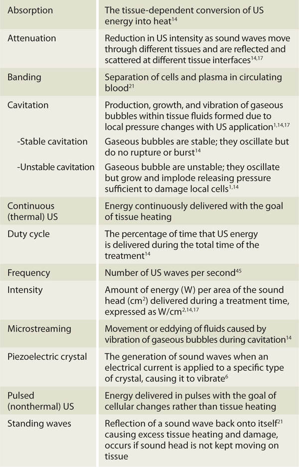
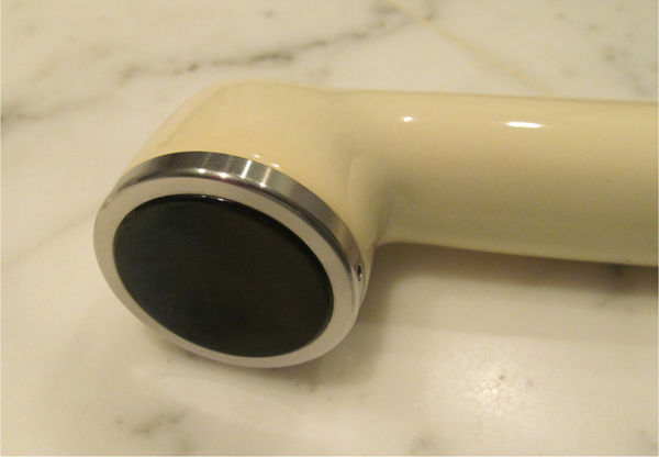
FIGURE 16-3 Ultrasound transducer that houses the piezoelectric crystal The ultrasound transducer or “head” houses the piezoelectric crystal that transforms the electricity into sound waves and is available in different sizes and shapes, depending on the purpose of the procedure.
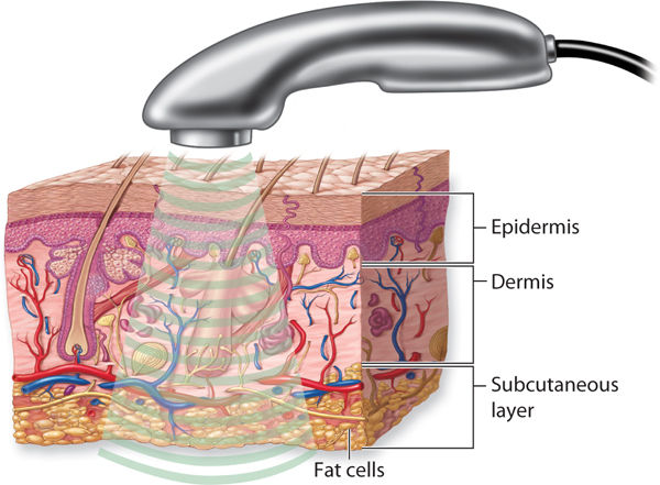
FIGURE 16-4 Transmission of sound waves Electric energy causes the piezoelectric crystal to vibrate and create sound waves that are transmitted to the dermal/subdermal tissue for wound healing.
US energy is utilized in wound healing due to its ability to effect changes in cellular activity. When therapeutic levels of sound waves come into contact with tissues, two primary mechanical processes are produced: cavitation and microstreaming.20 Acoustic cavitation refers to the generation of vibrating, microscopic bubbles in interstitial spaces.14,21 Bubble oscillations create compressive forces on surrounding cells as they move through tissue fluids, thereby stimulating changes in cell membrane activity.20 Cavitation is said to be “stable” when oscillating bubbles change in size but do not burst.14 Unstable cavitation occurs when the oscillating bubbles grow in size and implode, thereby causing free radical (OH−, H+) formation. Unstable cavitation increases tissue temperature and pressure, which can cause local tissue damage (FIGURES 16-5, 16-6).14
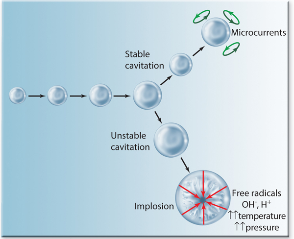
FIGURE 16-5 Illustration of microstreaming, stable cavitation, and unstable cavitation
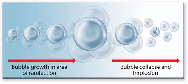
FIGURE 16-6 Unstable cavitation occurs when the transient gaseous bubbles implode, causing tissue damage and free radical formation. This process may assist in the breakdown of necrotic tissue.
Current eddies created in fluids by the oscillating bubbles produce unidirectional movement of tissue fluids thereby potentiating mechanical stimulation to surrounding cells.20,21 This type of fluid movement is referred to as microstreaming. US-generated stable cavitation and microstreaming have been shown to produce changes in cellular diffusion and growth factor production.17,20
EFFECTS AT THE CELLULAR AND TISSUE LEVELS
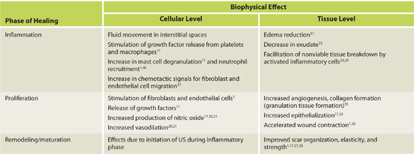
US has the potential to positively affect tissue during all phases of wound healing. Pulsed US does not generate heat, allowing for safe use during the acute inflammatory phase. The properties of cavitation and microstreaming change membrane permeability, thereby facilitating the release of growth factors, enzymes, and chemotactic agents.1,17,22 Facilitation of wound debridement also occurs by activation of the inflammatory cells involved in autolysis of nonviable tissues.23,24 In the inflammatory phase, this can cause earlier migration of proliferative cells, thereby hastening the onset of angiogenesis.17 In the proliferative phase, studies support that therapeutic US can accelerate granulation tissue formation and dermal repair by increased migration and activity of fibroblasts and endothelial cells.1,25,26 The positive effects of US seen during remodeling (also referred to as maturation) occur primarily due to early initiation of US during the acute inflammatory phase of healing. Published data document stronger, more organized, and more elastic scar tissue1,17,27,28 with early use of US and suggest increased collagen production as the likely mechanism of action. Since bacteria are present during all phases of wound healing, the role of US in reducing bioburden applies throughout the entire healing process. TABLE 16-3 lists the primary biophysical effects of US during inflammation, proliferation, and remodeling/maturation.
TABLE 16-3 Cellular and Tissue Biophysical Effects of Ultrasound by Phase of Healing
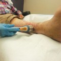
US energy can be delivered at either high or low frequency; TABLE 16-4 highlights clinically relevant differences between the two frequencies. High-frequency US (HFUS) devices traditionally used by physical therapists for tissue heating and repair require contact between the transducer and body tissue using a coupling medium (eg, US gel, water) to decrease reflection of sound waves.6 Cameron14 documents significant reflection of US energy by air and less than 1% reflection with the use of a suitable coupling medium. Low-frequency, noncontact US utilizes a fine saline mist as both a coupling medium and a mechanism of US energy transference (FIGURE 16-7).23
TABLE 16-4 Comparison between High- and Low-Frequency Ultrasound
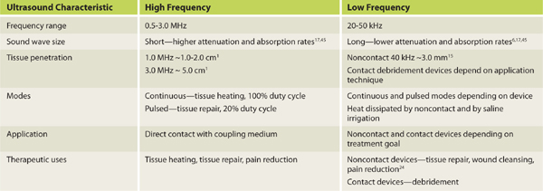
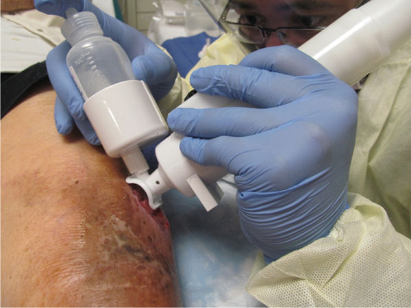
FIGURE 16-7 The bottle of saline attached to the disposable treatment applicator provides the fine saline mist that serves as the conducting medium for low-frequency, noncontact ultrasound
High-Frequency US
When HFUS is delivered in continuous mode at 1 to 3 megahertz (MHz), cavitation and microstreaming occur along with the thermal effect of tissue heating (FIGURE 16-8). Tissue heating occurs due to increased cellular vibration and frictional forces caused by continuous exposure to HFUS energy. Though rarely used in wound management,20 tissue heating can increase periwound tissue temperatures and result in increased local vasodilation and blood flow to the wound area.1,7 It has been documented that increasing periwound temperatures may reduce wound pain, although the exact mechanism of how this occurs is unclear.1 It is important to note that HFUS delivered in the continuous mode has the potential to be destructive and burns may occur in treated areas with insufficient blood flow to dissipate heat (FIGURE 16-9). A full list of precautions and contraindications associated with US is included in this chapter.
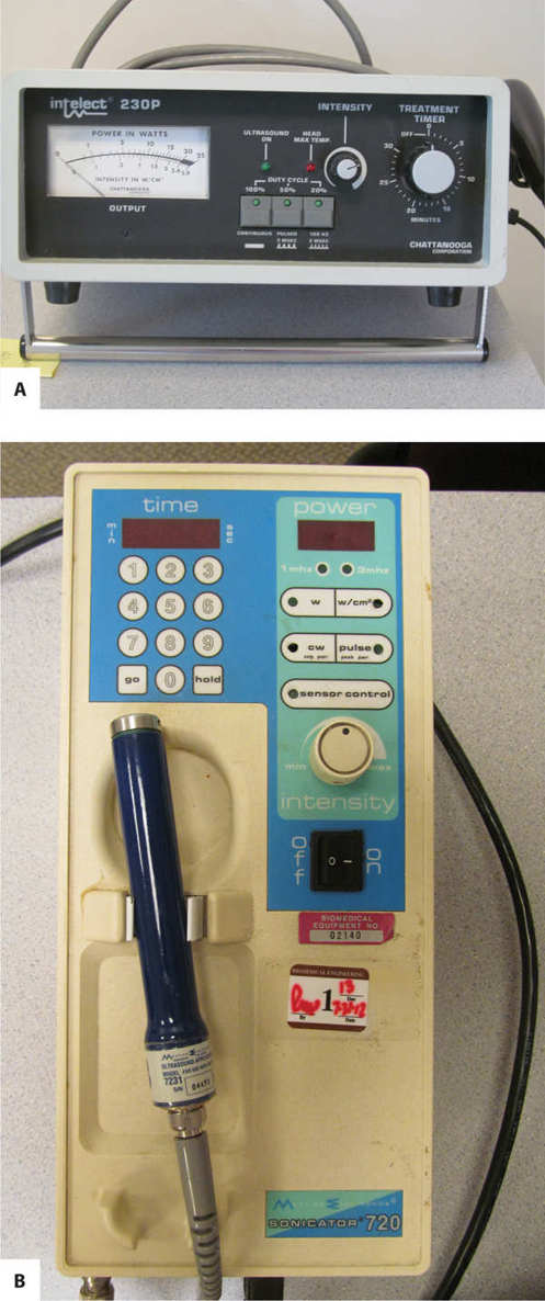
FIGURE 16-8 A, B Two traditional machines that provide high-frequency ultrasound.
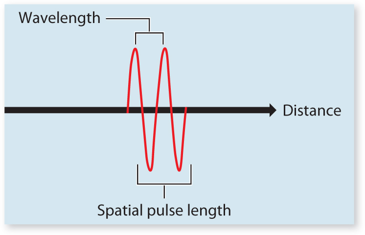
FIGURE 16-9 Continuous mode of delivery Ultrasound waves delivered in the continuous mode would continue along the distance, or the duration of treatment.
HFUS in pulsed mode produces the same cavitational and microstreaming effects in tissue as continuous mode but without the risk of tissue heating. Like continuous mode, pulsed mode can penetrate tissue up to 5 cm beneath the skin. In pulsed (nonthermal) mode, US energy is delivered in short pulses generating only small amounts of heat that is quickly dissipated resulting in no appreciable increase in tissue temperature. The benefits of pulsed mode US make it a safer and more effective form of therapeutic US delivery for wound management than continuous mode (FIGURE 16-10).1
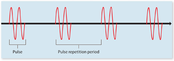
FIGURE 16-10 Pulsed mode of delivery Ultrasound waves delivered in the pulsed mode. No heat is generated in the tissue, and the sound waves can penetrate up to 5 cm beneath the skin.
Low-frequency US
Noncontact Low-frequency, noncontact US has become the primary method of US delivery for wound management (FIGURE 16-11). The MIST Therapy device (Celleration Inc, Eden Prairie, MN) utilizes a thin mist of sterile normal saline solution23 as a coupling medium to transfer US energy to body tissues, thereby negating the need for the US head to make contact with wound tissues.15 MIST therapy delivers continuous US energy that does not result in tissue heating due to a low-intensity therapeutic treatment range, heat dissipating saline mist, and noncontact method of delivery.17
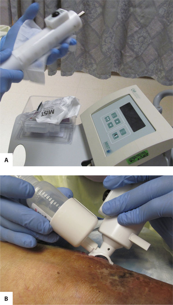
FIGURE 16-11 Noncontact, low-frequency ultrasound A. The noncontact, low-frequency ultrasound machine with a 1 cm2 sound head and disposable kit for delivery. B. The sound head is protected from touching the wound by the disposable cover that also holds the bottle of saline solution used as the conducting medium.
Low-frequency ultrasound (LFUS) produces higher levels of cavitation compared to HFUS, which allows low-frequency devices to deliver greater amounts of US energy to tissues during comparable treatment times.1 Using low-frequency (40 kHz) and low therapeutic intensities (0.1-0.5 W/cm2),15,17,23,24 MIST therapy has been shown to promote wound healing through tissue stimulation, promotion of fibrinolysis,1,15,25 and the removal of wound exudates and bacteria with thorough wound cleansing.23,24,29 Noncontact US may also play a role in active bacterial killing with damage to bacterial cell walls being the likely mechanism of action (FIGURE 16-12).15,16 It has been proposed that LFUS may alter the genetic code of certain bacteria, thus increasing antibiotic susceptibility and decreasing virulence.16 Additionally, this treatment modality is nontoxic and does not promote bacterial resistance.15,23 Studies support that using noncontact LFUS therapy as an adjunct to conventional wound management can result in faster reductions in wound surface area, decreased bioburden, and reduced pain (TABLE 16-5).15,19,24,25
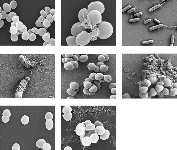
FIGURE 16-12 Treatment of bacteria with low-frequency, noncontact ultrasound Photographs showing the effect of low-frequency, noncontact ultrasound on the cell membrane of bacteria that commonly cause wound infections. (Kavros SJ, Schenck EC. Use of noncontact low frequency ultrasound in the treatment of chronic foot and leg ulcerations: a 51 patient analysis. J Am Pod Med Assn. 2007;97(2):95-101. Used with permission.)
TABLE 16-5 Benefits of Low-Frequency Ultrasound
Contact In wound management, low-frequency, contact US devices are primarily used for debridement of nonviable tissue. These devices produce highly focused ultrasonic energy that fragments or liquefies nonviable tissue using the principle of unstable cavitation as the mechanism of action.1 Portable devices such as the Qoustic Wound Therapy System (Arobella Medical LLC, Minnetonka, MN), Sonoca 180 (Soring Medical Technology, Doral, FL), and the SonicOne (Misonix, Inc, Farmingdale, NY) provide efficient, relatively painless,1 selective debridement in inpatient and outpatient settings.17
The US energy delivered by low-frequency contact devices may be in a continuous or pulsed mode depending on the specific manufacturer.17
Related posts:
Stay updated, free articles. Join our Telegram channel

Full access? Get Clinical Tree


