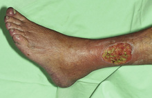
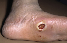
Pressure sites (lateral malleolar region)
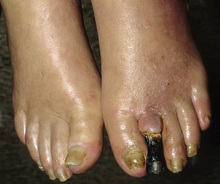
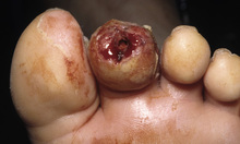
Distal points (toes)
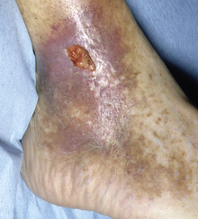
Irregular borders
Yellow fibrinous base
Dry, necrotic base
Well-demarcated (“punched out”)
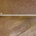
Yellow–brown to brown discoloration due to hemosiderin deposits
Pinpoint petechiae (“stasis purpura”)
Lipodermatosclerosis
Varicosities
Leg/ankle edema
± Stasis dermatitis
± Lymphedema
Weak/absent peripheral pulses
Prolonged capillary refill time (>3–4 seconds)
Pallor on leg elevation (45° for 1 min)
Dependent rubor
Peripheral neuropathy with decreased sensation
± Foot deformities
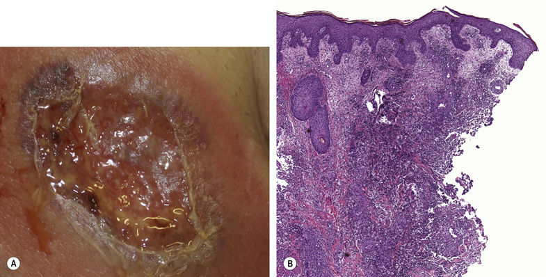
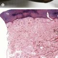
Fig. 17.1 Pyoderma gangrenosum. A Classic ulcerative pyoderma gangrenosum. The edge of this ulceration on the shin is undermined with a violet–gray color as well as an inflammatory rim. Note the central scarring. B In expanding untreated lesions, a diffuse infiltrate of neutrophils is present. A, Courtesy, Yale Dermatology Residents’ Slide Collection.
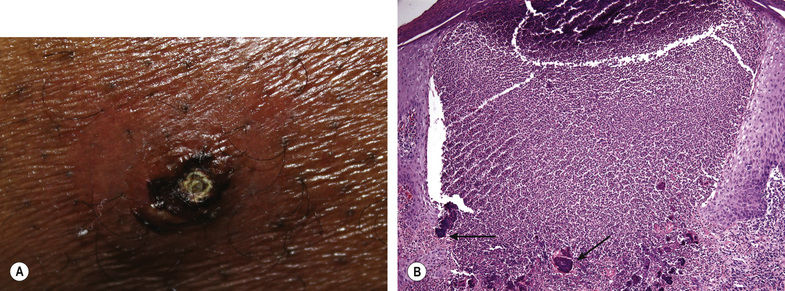
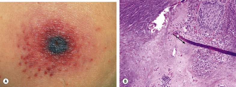
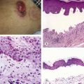
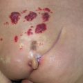
Fig. 17.3 Ecthyma gangrenosum. A Embolic lesion of Pseudomonas aeruginosa on the chest. Note the necrotic center and inflammatory border. B Histopathologic findings include dermal necrosis and a light blue haze of organisms (arrow). A, Courtesy, Yale Dermatology Residents’ Slide Collection.
Related posts:

Stay updated, free articles. Join our Telegram channel

Full access? Get Clinical Tree


