Body Site/Regional
Key Differences 
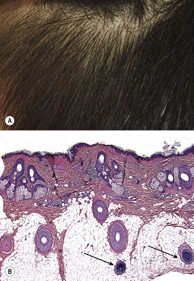
• Scalp – numerous anagen hair follicles with bulbs (arrows) in the fat (Fig. 1.1)
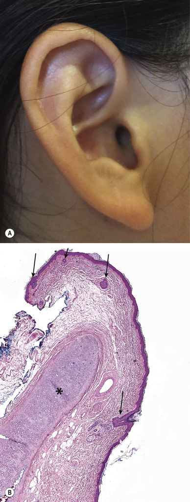
• Ear – vellus hair follicles (arrows) and central cartilage (*) (Fig. 1.2)
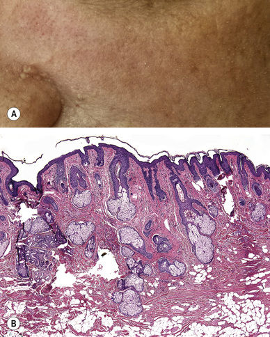
• Face – prominent hair follicles and sebaceous glands within the dermis (Fig. 1.3)
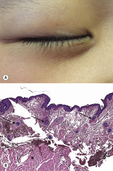
• Eyelid – vellus hair follicles (arrows) and underlying skeletal muscle (*) (Fig. 1.4)
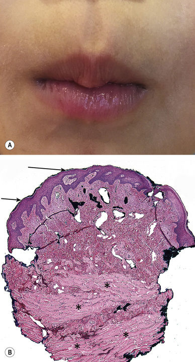
• Cutaneous lip – epidermis with keratin and a granular layer (arrows); skeletal muscle (*) (Fig. 1.5)
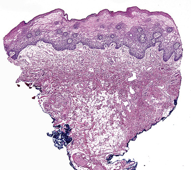
• Mucosal lip – pale epithelium that lacks a granular layer and does not keratinize (Fig. 1.6)
• Areola – acanthotic, pigmented epidermis with dermal smooth muscle bundles (arrows) (Fig. 1.7)
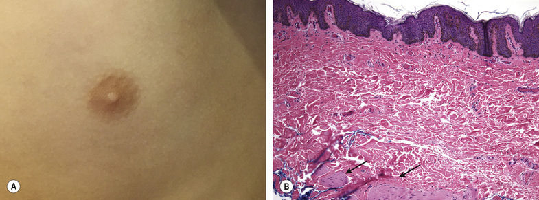
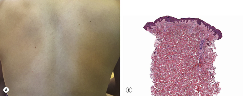
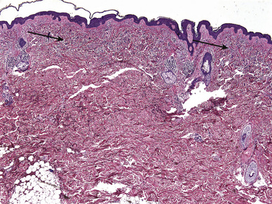
• Back – thick dermis (Fig. 1.8)
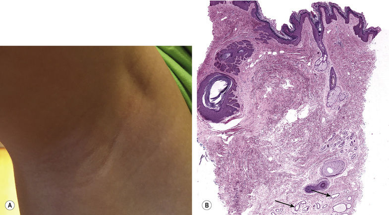
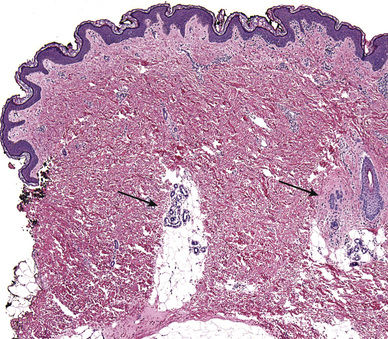
• Axilla – undulating epidermis with deep apocrine glands (arrows) (Fig. 1.9)
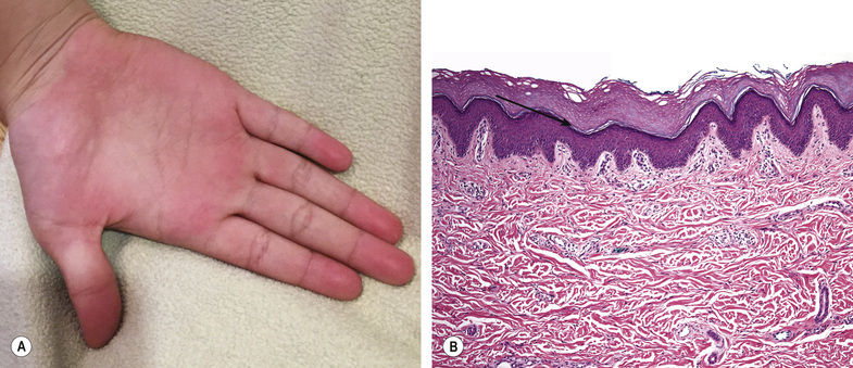
• Acral – thick stratum corneum with stratum lucidum (arrow) (Fig. 1.10)
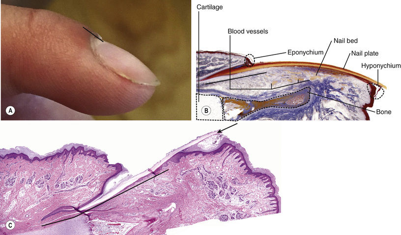
• Nail – nail plate (arrow), nail matrix (black bar), and nail bed (Fig. 1.11)

Distribution
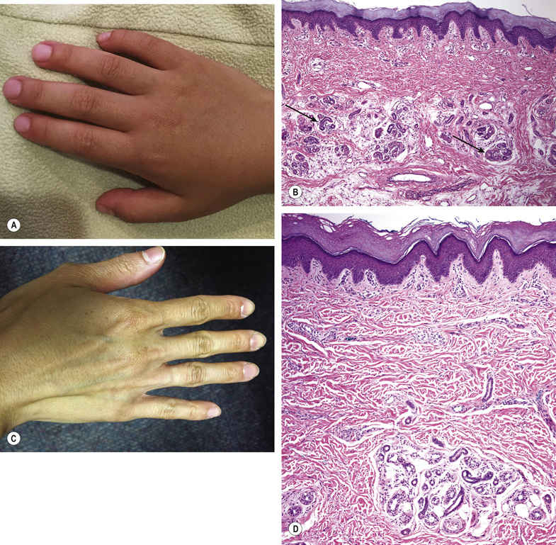
Extensive – more than one body part affected with multiple lesions; may preferentially affect extensor surfaces (e.g. elbows/knees) vs flexor surfaces (e.g. antecubital/popliteal fossae) or ventral vs dorsal surfaces (Fig. 1.15)
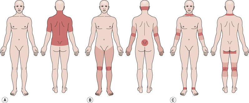
Photodistribution – can vary depending on the type of clothing worn; sun-protected sites of the face/neck generally include the central upper cutaneous lip and submental area (Fig. 1.16A; see Fig. 3.7)
Double-covered areas – generally includes sites covered by undergarments (Fig. 1.16B)
Acral – hands/feet but also the tips of the ears/nose (Fig. 1.16C)
Body folds – inframammary/intertriginous (Fig. 1.16D)
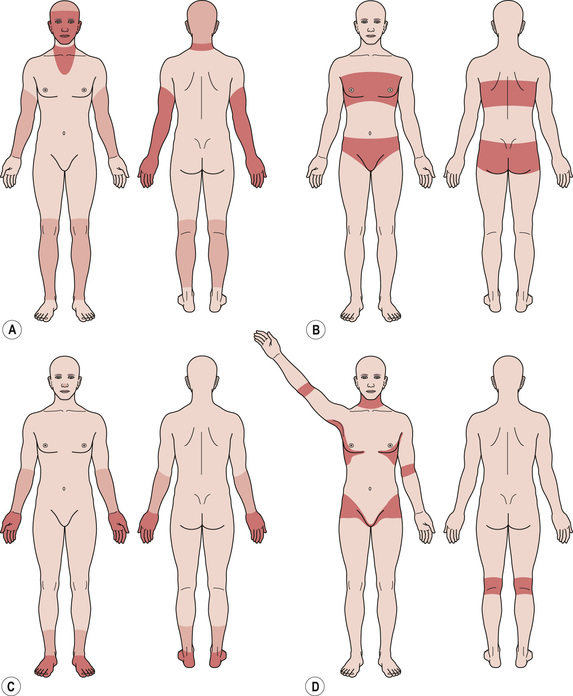
Generalized – involving the majority of the cutaneous surface (Fig. 1.17)
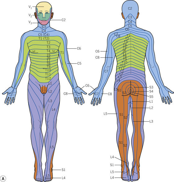
Dermatomal – patterns of cutaneous innervation by spinal nerve roots (Fig. 1.18A)
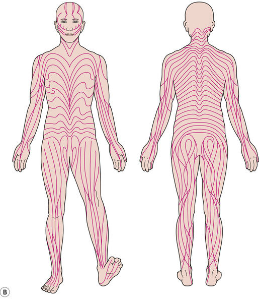
Blaschkoid – follows patterns of embryonic cell migration; while the linear pattern on the extremities is similar to the dermatomal pattern, the V-shaped curves over the trunk and the S-shapes on the abdomen are characteristic (Fig. 1.18B)
Patterns
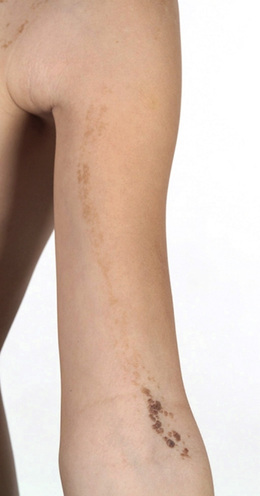
Thin stripes – often corresponds to Blaschko’s lines (see Fig. 1.18B, Fig. 1.19)
Related posts:

Stay updated, free articles. Join our Telegram channel

Full access? Get Clinical Tree


