Absent comedones
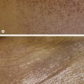
Benign Cephalic Histiocytosis (Fig. 15.2)
Typically on the face or neck
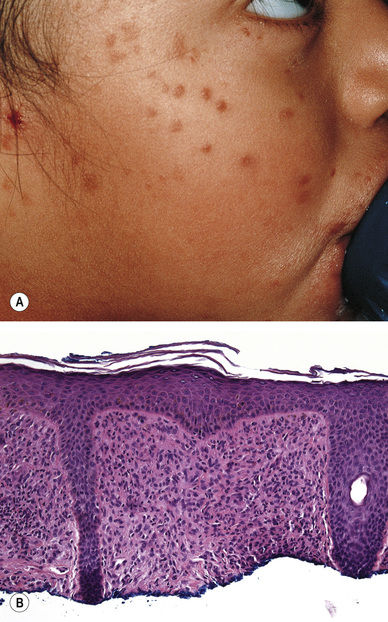
Small (<5 mm) brown–red papules
Spontaneous resolution over time (months to years)
Histopathology:
Histiocytes (CD1a-negative) within the dermis
Body Folds
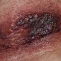
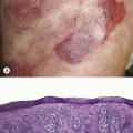
Langerhans Cell Histiocytosis (Fig. 15.3)
Favors the scalp and body folds
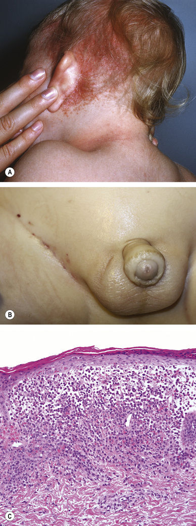
Pink to red–brown papules, often with petechiae, sometimes eroded/ulcerated
Histopathology:
Histiocytes with reniform (kidney-shaped) nuclei that are langerin-, CD1a-, S100-positive
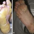
Acral
Acropustulosis of Infancy (Fig. 15.4)
Cyclical, typical age is 3 to 6 months up to 2–3 years of age
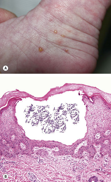
Pruritic vesicles
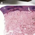
Histopathology:
Intraepidermal pustules
Scabies (Fig. 15.5, See Fig. 7.16A,B)
Can present like acropustulosis of infancy
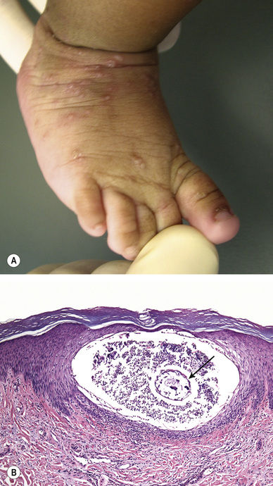
Histopathology:
Evidence of scabies (mite – arrow) infestation on scraping or biopsy
Related posts:

Stay updated, free articles. Join our Telegram channel

Full access? Get Clinical Tree


