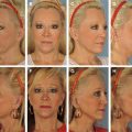
Aging of the midface is a complex aesthetic problem requiring an individualized and multifaceted surgical approach. The objective of harmonious rejuvenation of the entire face as well as increasing patient interest in midface rejuvenation mandates surgical familiarity with these techniques. Midface rejuvenation procedures have evolved from traditional laterally based rhytidectomy techniques with superolateral elevation to modern centrofacial approaches designed to achieve more vertical vectors of elevation. These approaches are informed by an evolving understanding of the multiple processes that contribute to the aged appearance of the midface and are based on lower blepharoplasty surgical techniques.
Key points
- •
Midface rejuvenation procedures have evolved from laterally based rhytidectomy techniques to centrofacial approaches with more vertical vectors of elevation.
- •
Volume loss and remodeling of the maxillofacial skeleton should be addressed with various augmentation procedures.
- •
Patients with prominent globe/negative vectors or previous lower eyelid surgery are at increased risk for postoperative lid malposition.
- •
Lateral canthal support procedures range from simple midface elevation and suspension to formal canthotomy/cantholysis with lateral canthal reconstruction; selecting the appropriate technique should be guided by individual patient concerns and surgeon experience and skill.
- •
The tear trough and nasolabial fold can be difficult areas to correct surgically; fillers, autologous free fat or superficial musculoaponeurotic system (SMAS) grafts, or allografts can be transferred to the tear trough area or nasolabial folds.
The midface is an important facial aesthetic subunit and may exhibit many of the telltale signs of aging. Aging occurs within each of the anatomic layers of the midface, with changes visible as early as the third decade. Descent of atrophic skin and attenuation of orbicularis musculature are often associated with pseudoherniation of orbital fat, outwardly transforming the smooth concavity of the eyelid-cheek junction into a double convexity. Tarsoligamentous laxity leads to visible lengthening of the lower lid, and the eye assumes a rounded shape with a negative canthal tilt. Crow’s-feet form at the superolateral boundary of the midface. Soft tissues over the malar eminence, in particular the subcutaneous malar fat pad and midface retaining ligaments, become ptotic and descend, reducing malar prominence and skeletonizing the inferior orbital rim, creating a hollowed appearance. Malar bags and festoons manifest within the superficial layers. A nasojugal trough becomes apparent medially, resulting in the so-called tear trough deformity. The dependent region of the nasolabial crease becomes burdened with ptotic superolateral soft tissues, giving rise to a hooded appearance and a deepening of the fold. Volume loss has been increasingly appreciated as an important part of the aging process as well progressive remodeling of the maxillofacial skeleton. The skin of the face undergoes typical actinic processes. The sum of these changes leads to the characteristically tired appearance associated with age, with the overall loss of the heart-shaped face of youth. A thorough understanding of the anatomy of the midface and its age-associated pathophysiology is essential to the safe and effective performance of midface rejuvenation.
Anatomy and pathophysiology
Surgical midface anatomy is complex. Skin, various fat pads, the SMAS, mimetic musculature, neurovascular structures, retaining ligaments, and periosteum overlie the facial skeleton in a layered fashion. Critical structures pertaining to transpalpebral facelift techniques include the lower lid skin, orbital septum, lateral canthal tendon, orbicularis oculi muscle, suborbicularis oculi fat (SOOF) and malar fat pads, and zygomatic and orbicularis retaining ligaments.
The anatomy of the orbicularis oculi is central to discussion of transpalpebral techniques. It is a facial mimetic muscle and an integral part of the SMAS, functioning as the ocular sphincter. It is composed of a central pars palpebralis and a peripheral pars orbitalis, and, together with the lower eyelid skin, comprises the anterior lamellae of the eyelid. Its orbital part underlies the subcutaneous malar fat pad as it radiates inferiorly across the lid-cheek junction, whereas the SOOF pad lies deep to the muscle and generates a natural glide plane that allows the muscle to move independently over the underlying periosteum and origins of the lip elevator muscles. This plane has been described in thoughtful anatomic study as the prezygomatic space. The orbicularis retaining ligament attaches the deep aspect of the orbicularis muscle to the inferolateral orbital rim periosteum, forming the superior boundary of the prezygomatic space and separating it from the preseptal space of the lower eyelid. This ligament has also been interpreted as an orbitomalar ligament, inserting into the dermis through the muscle body; regardless, release or elevation of this ligament is fundamental to achieving satisfactory elevation of deep midface tissues. Laterally, this ligament expands and joins the lateral orbital thickening, which overlies the bony inferolateral orbital rim and represents a confluence of the major superficial and deep fascias of the lateral orbital and temporal regions. Inferiorly, zygomatico-cutaneous ligaments arise between the zygomaticus muscles to fix the malar fat pad and cheek skin to zygomatic eminence. These are also important retaining ligaments of the midface that must be released to elevate deep tissues and define the inferior extent of the prezygomatic space (discussed previously). The lateral canthal tendon is a critical support structure that anchors the lateral canthus to the bony orbit and is contiguous with the superficial fascial component that joins the lateral orbital thickening. It defines the lateral canthus of the eye and damage to or improper reconstruction of the lateral canthal tendon can lead to significant cosmetic deformity. Inferiorly, the nasolabial fold represents an area of confluence of tissues at the lower border of the midface, where fasciofibrous connections of the SMAS and mimetic musculature join with the dermis.
Transpalpebral approaches
Hester and colleagues are credited with the introduction of the skin-muscle flap approach to the midface in 1996. Their technique demonstrated the importance of proactive lower lid support and showed that multiple points of reliable suture fixation are essential to achieving stable fixation. They also emphasized conservative skin resection and defined the major tenets of the transblepharoplasty midface approaches. In 1999, Gunter and Hackney modified this procedure to avoid requisite lateral canthoplasty, pioneering a different approach the management of the lateral canthus.
Access to the midface is typically subciliary, as in Hester and colleagues’ original technique, or transconjunctival. The former offers the ability to perform conservative skin and muscle resection and affords wider exposure to the midface but carries a theoretic risk of microdenervation of the pretarsal orbicularis muscle. The latter preserves orbicularis innervation and minimizes septal manipulation that may lead to contracture and lid retraction but has more limited exposure (which may be improved by adding a canthotomy). Patients with significant malar bags or festoons may benefit from a traditional skin-muscle flap technique, because these superficial deformities are unlikely to be corrected via a transconjunctival approach.
Key considerations in lower blepharoplasty
Preoperative assessment should include an ophthalmic history and evaluation. Dry eyes, prior procedures, glaucoma, and inflammatory conditions should be noted. Patients should be counseled that patients prone to dry eyes tend to have worsening of symptoms in the postoperative period and the need for ocular lubricants should be discussed. Surgery may not be appropriate for some of these patients. Of critical importance to aesthetic surgeons and patients is avoiding malposition of the lower lid and lateral canthus. Patients with prominent globe or negative vector, in whom the plane of the cornea is anterior to the plane of the bony orbital rim, as well those who have undergone lower lid surgery are at increased risk for postoperative lid malposition. Preoperative or intraoperative distraction and snap tests should be performed to assess lid laxity and further discern the need for conservative anterior lamella resection or anticipated need for lower lid support.
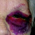
Pseudoherniation of orbital fat is well known to give rise to contour irregularities of the lower eyelid. In midface surgery, this particular deformity requires additional consideration and must be dealt with in a thoughtful manner. Conservative resection of lower lid fat compartments can be performed during a transblepharoplasty midface lift, with limited resection of fat, particularly in the nasal and midfat compartments. Temporal fat tends to be more forgiving and may be excised more aggressively, which is done routinely by some surgeons. The need for conservative resection of fat should be carefully assessed preoperatively and intraoperatively once the flap is elevated. After release of the arcus marginalis, infraorbital fat can be used to redrape the skeletonized inferior orbital rim and fill in the nasojugal trough. Elevation of the midface soft tissues with arcus marginalis release may provide adequate correction of those irregularities preoperatively attributed to pseudoherniation, and overaggressive fat excision, therefore, may lead to lower lid complications.
Technique
The specific techniques described represent those preferred by the senior author (APS). These described procedures nonetheless illustrates the essential techniques of transpalpebral midface rejuvenation ( [CR] ).
The transpalpebral midface lift can be performed under intravenous sedation or general anesthesia and with the subciliary approach in carefully selected patients, even under local anesthesia. Careful regional nerve blockade should be applied, and local anesthesia can be infiltrated along planned incision lines.
Both subciliary and transconjunctival incisions can be used to perform a transpalpebral midface lift, depending on the anticipated extent of lower lid skin resection. Preoperative evaluation of lower lid laxity with snap and distraction tests aids in this determination.
In patients who do not have significant lower lid skin redundancy, a transconjunctival incision with canthotomy and cantholysis is performed. A small incision is made from the lateral canthus extending to a point 4 to 6 mm laterally within an existing crow’s-feet rhytid. A straight clamp is introduced straddling the lateral canthal conjunctiva and used to crush the soft tissue to limit bleeding, and the incision is carried down to bone using electrocautery. Cantholysis is carried out using curved Stevens scissors, directed inferiorly to divide the inferior limb of the lateral canthal tendon.
The transconjunctival incision is then made approximately 3 mm below the inferior tarsal border and is carried laterally to join the canthotomy incision. Dissection proceeds inferiorly in a preseptal plane, separating the conjoined capsulopalpebral fascia and orbital septum. This plane is followed to the inferior orbital rim, where the periosteum is incised with preservation of a 3- to 4-mm cuff ( Fig. 1 ). This marks the entry point to the subperiosteal plane. The conjunctival flap is draped over the cornea for protection.
If preoperative examination reveals excessive lid skin redundancy, a subciliary incision is utilized after canthotomy/cantholysis. The excision is extended laterally to join the crow’s-feet incision. A suborbicularis plane is developed by dividing the muscle at an angle, preserving a cuff of pretarsal orbicularis. The skin-muscle flap is elevated to expose the middle lamella or orbital septum ( Fig. 2 ). Dissection proceeds inferiorly to the inferior orbital rim, and the periosteum is incised (in the manner described previously) to access the subperiosteal plane.
The periosteum is elevated off the facial skeleton. Dissection boundaries are the gingivobuccal sulcus inferiorly, the malar eminence and medial insertion of the masseter laterally, and the nasomaxillary junction and pyriform aperture medially. Care should be taken to preserve the infraorbital ( Fig. 3 ) and zygomaticofacial ( Fig. 4 ) nerves as they exit their respective foramina. The infraorbital nerve should be completely skeletonized (see Fig. 3 ) and the orbital retaining ligament should be appropriately released and elevated to aid in mobilizing the midfacial tissues. An inferior periosteal incision is created near the gingivobuccal sulcus, and a clamp can be used to spread until superficial fat can be seen through the incision. This marks the full mobilization of the midface composite flap ( Fig. 5 ), which, with superior traction, should be easily elevated without excessive force. To ensure proper mobility of the flap, lateral soft tissues should be swept away until the masseter origin is clearly visible.