The eyes play a central role in the perception of facial beauty. The goal of periorbital rejuvenation surgery is to restore youthful proportions and focus attention on the eyes. Blepharoplasty is the third most common cosmetic procedure performed today. Because of the attention placed on the periorbital region, preventing and managing complications is important. Obtaining a thorough preoperative history and physical examination can significantly reduce the incidence of many of the complications. This article focuses on the preoperative evaluation as it relates to preventable complications, followed by common intraoperative and postoperative complications and their management.
Key points
- •
Most complications can be prevented by proper preoperative history and physical examination.
- •
A thorough understanding of the complex periorbital anatomy prevents inadvertent injury to crucial structures.
- •
Many complications can require conservative management with topical lubrication and waiting the appropriate time before performing any further intervention.
- •
Severe complications such as visual loss from orbital hemorrhage require prompt identification and treatment.
- •
Midface surgery is a safe and well-tolerated procedure, with most complications arising from imprecise implant, suture, or endotine placement.
Introduction
The eyes play a central role in the perception of facial beauty. The goal of periorbital rejuvenation surgery is to restore youthful proportions and focus attention on the eyes. This is validated by the public’s interest in blepharoplasty, which is the third most common cosmetic procedure performed today, and for the foreseeable future. Because of the attention placed on the periorbital region, the importance of preventing and managing complications in this area cannot be overstated. Critical to this concept is obtaining a thorough preoperative history and physical examination, which can reduce significantly the incidence of many complications. This article focuses on the preoperative evaluation as it relates to preventable complications, followed by common intraoperative and postoperative complications and their management.
Preoperative evaluation for periorbital and midface rejuvenation surgery
After discussing the patient’s goals of surgery, obtaining a comprehensive medical and surgical history is critical to preventing complications from periorbital and midface surgery. Patients should provide a detailed list of all prescription and nonprescription medications, including topical eye drops. In additional to a general medical history evaluation, special attention should be placed on prior facial surgeries, history or symptoms of coagulopathies, prior anesthesia reactions, and conditions that may alter healing, such as diabetes and immunosuppression. Patients with systemic diseases such as Sjögren syndrome, rheumatoid arthritis, Grave’s disease, or neuromuscular diseases should be evaluated appropriately and counseled regarding the increased risks.
Several aspects of the patient’s history are specific to preventing complications of periorbital surgery. Keratoconjunctivitis sicca, or dry eye syndrome (DES) is a common condition that has a wide range of etiologies. Patients should be questioned regarding symptoms of dry eyes (redness, soreness, mucoid discharge changes), because blepharoplasty can both result in DES, or exacerbate the condition if present before surgery. Prischmann and colleagues reviewed 892 cases of both lower and upper blepharoplasty and evaluated the results of a dry eye questionnaire that was given to patients before and after their surgery. They found the incidence of DES and chemosis was 26.5% and 26.3%, respectively. Several variables also increased the incidence of both DES and chemosis, including concurrent upper and lower blepharoplasty, transcutaneous approaches, preoperative DES, preoperative skin laxity, male sex, and hormone therapy. They recommended that, in patients with increased risk factors, adjunctive lid tightening procedures should be performed to reduce the risk of this complication.
Korn and colleagues reported on the correlation between laser in situ keratomileusis (LASIK) and blepharoplasty and found that all 6 patients in the study developed DES. Special precautions must be taken in these patients to prevent this complication.
During the preoperative evaluation, it is important to identity patients with Grave’s disease. This condition is owing to autoimmune activity against thyroid-stimulating hormone receptors and is associated with orbital disease in 40% of patients. It is characterized by glycosaminoglycan deposition, fibrosis of extraocular muscles, and adipogenesis in the orbit. Patients suspected of having this condition should be evaluated by an appropriate specialist to manage and prevent medical complications. Although blepharoplasty is often required in the surgical management of this disease, there are often multiple stages and varying techniques to address the proptosis and lid retraction that are associated with this condition.
A thorough periorbital examination is required before undertaking any operation to properly identify the anatomic structures contributing to the patient’s complaints and to recognize features that can increase the risk of postoperative complications. Critical to maximizing outcomes after upper blepharoplasty is preoperative evaluation of the brow. Excessive contraction of the frontalis muscle occurs to compensate for significant upper lid dermatochalasia. If blepharoplasty is performed alone, relaxation of the brow will occur after surgery, which reduces the effectiveness of the operation ( Fig. 1 ). To properly identify brow ptosis, evaluate the patient in a relaxed gaze, or manually fixate the brow digitally while assessing for excess skin.
Failure to recognize upper eyelid ptosis results similarly in a dissatisfied patient. Ptosis is defined as a margin-to-reflex-1 distance of 2.5 mm or less. Proper preoperative examination and documentation is critical, because traditional upper blepharoplasty techniques alone does not address this condition adequately. In addition, ptosis is a potential complication of the surgery, and the etiology can be unclear if preoperative documentation omitted this physical examination finding.
Preoperative assessment of the upper eyelid crease is essential for incision planning. Unusually high creases can be a sign of levator dehiscence that, if present, should be addressed at the time of surgery. Lacrimal gland prolapse can masquerade as excessive fat, and injury to this structure can lead to postoperative complications such as DES ( Fig. 2 ). The lacrimal gland is located laterally and intraoperatively has a pinkish color. If significant prolapse is present, this can be easily managed by pexying the gland to the orbital rim.
Proper evaluation of the lower lid helps to prevent complications from lower blepharoplasty such as ectropion/entropion, lid malposition, DES, and chemosis. Horizontal lid laxity is measured by the snap test, in which traction is place on the lid and abruptly released ( Fig. 3 ). Prompt return to the globe indicates proper tone. The distraction test also measures lid laxity by maximally pulling the lid anteriorly. A distance of 6 to 8 mm indicates excessive lid laxity, which should be addressed by either lid tightening or shortening techniques. Malposition of the lower lid should also be assessed by placing upward traction on the lower lid. A normal lid should elevate to at least the midpupillary level. This evaluation is particularly important in revision cases.
Preoperative evaluation of excess skin in the lower eyelid is essential to prevent anterior lamellar shortening. This is evaluated by having the patient open their mouth widely while pinching the lower eyelid skin. If there is no excess in this position, removal of skin will put the patient as risk for lower lid malposition and ectropion.
A negative vector eyelid is described as a prominent globe with a recessed orbital rim/maxilla ( Fig. 4 ). Blepharoplasty in these patients is associated with higher complication rates, particularly lid malposition. The Hertel exophthalmeter measures the distance from the most anterior point of the lateral orbital rim and the anterior border of the globe. The normal range for this test is 15 to 17 mm, with a range of 12 to 22 mm. For patients with significant exophthalmos, operative intervention should be modified by performing minimal fat excision or using spacer grafts to reduce the incidence of lid malposition. In addition, canthal-tightening procedures can exacerbate the malposition, as increased tension along the prominent globe will force the lid to retract further.
Because of the many surgical options to manage facial rejuvenation of the midface, the challenge lies in the proper evaluation and diagnosis of the specific anatomic changes that require correction. Patients should be questioned regarding prior history of midface trauma or rejuvenation techniques that have been utilized in the past. The entire lower-lid–cheek contour should be evaluated as a whole to maximize patient satisfaction. Evaluation and documentation of facial nerve function is important, because facial nerve injury is a potential complication. Patients with significant skin laxity and jowling are often better treated by rhytidectomy techniques either alone or in conjunction with midface lifting. Preoperative evaluation of the lateral canthus is also warranted; some authors have reported upward deflection after this surgery.
Complications from upper and lower blepharoplasty
Meticulous preoperative evaluation and planning are essential to maximize patient outcomes and minimize complications. However, even the most adept and thoughtful surgeons will have complications in their practice. When complications do occur, the goal is to rapidly identify them and quickly initiate the proper treatment to minimize morbidity. Intraoperative and postoperative complications and their management are summarized ( Table 1 ).
| Periorbital Complications | Cause | Prevention | Management |
|---|---|---|---|
| Dry eye syndrome | Missed preoperative, exposure, irritation | Preoperative history/examination, eye protection | Lubrication, lacrimal puncta ducts |
| Chemosis | Conjunctival irritation, disruption of lymphatics | Temporary tarsorrhaphy | Topical dexamethasone, topical phenylephrine, lubricants, conjunctivotomy and patching for severe cases |
| Ptosis | Missed preoperative, mechanical factors, levator injury | Preoperative history/examination, preserving levator | If persistent after 3 months, surgical ptosis repair |
| Diplopia/strabismus | Scar tissue, muscle injury | Limited deep cautery in lateral fat pad, muscle preservation | Local steroid injections, corrective strabismus surgery |
| Persistent dermatochalasia | Missed brow ptosis, underresection | Preoperative history/examination | Revision surgery, brow lift |
| Lower lid retraction/lagophthalmos | Muscle injury, preoperative lid laxity, anterior/posterior lamellar scarring, overzealous skin excision | Preoperative history/examination, canthal tightening procedures | Canthopexy/plasty, spacer graft, skin grafting, midface lifting |
| Corneal abrasion | Corneal exposure during surgery | Corneal protector | Lubrication, topical antibiotics |
| Hemorrhage | Anticoagulant use, vascular injury | Meticulous hemostasis | Surgical exploration, lateral canthotomy |
| Lacrimal gland injury | Missed preoperative, improper diagnosis | Proper identification intraoperatively | Lubrication |
| Infection | Surgical wound contamination, improper sterile technique | Perioperative antibiotics | Oral antibiotics, surgical decompression for abscess formation |
| Overcorrection | Inappropriate preoperative assessment | Thorough preoperative assessment, conservative fat removal | Postoperative fat grafting, fillers |
| Undercorrection | Inappropriate preoperative assessment | Thorough preoperative assessment, marking | Revision surgery |
| Midface | |||
| Sensory nerve damage | Surgical error | Subperiosteal dissection | Expectant management |
| Facial nerve damage | Cautery, wrong plane of dissection | Subperiosteal dissection | Expectant management |
| Infection | Oral contamination | Copious irrigation, possibly avoiding intraoral route | Antibiotics, surgical drainage as indication |
| Asymmetry | Suture fixation | — | Revision surgery |
| Implant malposition | Improper placement, shifting | Tight subperiosteal pocket, implant fixation | Revision surgery |
Intraoperative Complications
Corneal abrasions are a potential perioperative complication in any type of surgery. Inadvertent injury to the corneal epithelium can occur from oculotoxic sterilization chemicals, surgical instruments, gauze, or desiccation of the ocular surface. This complication is prevented by using corneal shields, which can be placed after applying tetracaine drops to the eye and a generous amount of petroleum-based lubricant. When corneal abrasions do occur, patients often complain of pain, foreign body sensation, burning, tearing, and blurry vision. Suspicion of this injury warrants prompt ophthalmologic evaluation with a fluorescein stain and a slit lamp. Management of these injuries depends on the severity, but most can be managed with 48 hours of topical erythromycin or bacitracin. The surface can also be protected with application of contact lens. Thermal injuries have been reported after cosmetic blepharoplasty and have resulted in postoperative astigamatism. Treatment consists of topical antimicrobials followed by topical steroids to reduce scar tissue formation. Persistence of the astigmatism may require corrective astigmatic keratomoty.
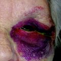
Intraoperative and postoperative hemorrhage is a potentially devastating complication owing to the potential risk of blindness. Some degree of ecchymosis is expected after the procedure; therefore, a thorough clinical evaluation is necessary to correctly screen for those who require urgent intervention. Retrobulbar hemorrhage leads to a compartment syndrome, which puts significant pressure on the intraorbital contents. The exact mechanism of blindness from retrobulbar hemorrhage has been attributed to either central retinal artery compression or direct optic nerve injury. Mejia and colleagues conducted a survey of 648 surgeons who performed 752,816 blepharoplasties, and found that the overall incidence of blindness from all causes was 0.0052%, and permanent blindness at 0.0033%. Hemorrhage can occur owing to vascular injury during injection, improper handling of orbital fat, and patient factors such as coagulopathies. Most instances occur within several hours after the procedure, but have also been reported several days after surgery.
Retrobulbar hematoma presents with rock hard proptosis, chemosis, severe pain, and visual changes ( Fig. 5 ). Ophthalmic consultation is requested early on in the course of therapy. Intraocular pressure is also elevated; however, surgical management is more dependent on the presence of visual changes. For patients with intact vision, conservative measures of cold compresses, intravenous osmotic agents, topical β-blocker drops, and acetazolamide are initiated. For patients with visual loss, aggressive management is required beginning with a lateral canthotomy. The wound should be opened to allow for decompression and washout. Any areas of active hemorrhage should be stopped with cautery. For patients with return of vision, they can be monitored closely and discharged with antibiotic prophylaxis and a tapered steroid dose pack. Persistent visual should be evaluated with an orbital CT. If a posterior hematoma is present, bony decompression is warranted.