TRAM
Peter Henderson
Jeffrey A. Ascherman
Joseph J. Disa
DEFINITION
The pedicle transverse rectus abdominis myocutaneous (TRAM) flap for breast reconstruction was first described by Hartrampf et al. in 1982, and it heralded the onset of a new era of abdomen-based autologous breast reconstruction. This procedure has been modified and improved upon, such that today free perforator flaps from the abdomen (as well as other sites such as the buttocks and thighs) have become the most modern, cutting-edge procedures available. But it is indisputable that the pedicle TRAM is a procedure that every reconstructive surgeon must have available in his or her armamentarium, to use as both a form of primary breast reconstruction as well as a salvage procedure in the case of failure of device-based reconstruction or other non-abdomen-based autologous reconstruction.1,2
ANATOMY
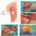
The rectus abdominis (RA) is a Mathes-Nahai type III muscle. The two dominant pedicles are the superior epigastric vessels (off the internal mammary artery and vein) and the deep inferior epigastric vessels (off of the external iliac artery and vein). The superior epigastric vessels approach the RA from the cranial direction on the central portion of the deep surface of the RA. The deep inferior epigastric vessels approach the RA from the lateral aspect of the caudal portion of the RA, on the deep surface (FIG 1).3
The origins of the RA are the sternum and the 5th-7th ribs.
The insertion is the pubic symphysis.
Innervation is from multiple branches of the intercostal nerves, which penetrate the lateral aspect of the posterior rectus sheath and enter the RA muscle on its deep surface.
The function of the paired RA is to flex the trunk at the waist.
The rectus sheath is composed of the aponeuroses of the external oblique, internal oblique, and transversus abdominis muscles (FIG 2). Cranial to the arcuate line (aka “arcuate line of Douglas” and “linea semicircularis”), the anterior rectus sheath is composed of the aponeuroses of the external oblique and the anterior leaflet of the internal oblique, and the posterior rectus sheath is composed of the aponeuroses of the posterior leaflet of the internal oblique and the transversus abdominis (as well as the transversalis fascia). Caudal to the arcuate line, the anterior rectus sheath is composed of the aponeuroses of the external oblique, internal oblique, and transversus abdominis muscles, and the posterior rectus sheath is composed of only the transversalis fascia.
PATHOGENESIS
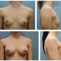
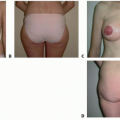
The pedicle TRAM procedure is indicated for breast reconstruction when skin and/or subcutaneous tissue is needed, and the patient has sufficient excess abdominal skin and fat to serve as donor tissue.
PATIENT HISTORY AND PHYSICAL FINDINGS
The patient’s breast history (prior procedures as well as adjuvant or neoadjuvant therapy) should be elicited to understand the timing and expected progression of the sequelae.
Nicotine use is highly deleterious to the outcomes of this operation and should be avoided at all costs. Tumor resection must occur in a timely fashion, but reconstruction can be delayed until the patient has stopped smoking.
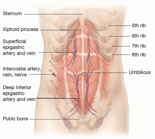
FIG 1 • Rectus abdominis anatomy.
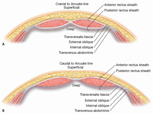
FIG 2 • Contributions to rectus sheath cranial and caudal to arcuate line.
In patients who will undergo radiation therapy, the surgeon and patient may choose to delay the reconstruction until the radiation is completed.
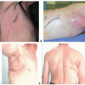
Surgical history should focus on prior abdominal operations, breast operations, and cardiac procedures (to determine if the internal mammary artery has been used as a cardiac bypass or has potentially been damaged during rewiring of a sternotomy; this is the source vessel for the superior epigastric artery).
Physical exam of the chest should focus on assessing the surface area of skin needed, and the volume of breast mound needed.
The abdomen should be examined for amount of subcutaneous tissue and excess skin and estimated laxity (in terms of defect that can be closed without undue tension). Prior abdominal scars should be noted (midline scars usually render the tissue contralateral to the pedicle unable to be included in the flap). Any hernias (umbilical, incisional, etc.) should be noted.
An acoustic (handheld) Doppler can be used to identify perforators (especially if a fascia-sparing approach is to be used)
IMAGING
CT angiography can be performed if there is any question about the patency of the superior epigastric vessels, as well as for identification of abdominal wall perforators.
NONOPERATIVE MANAGEMENT
Breast reconstruction is never mandatory and can be deferred if the patient’s wishes or comorbidities preclude it from happening.
SURGICAL MANAGEMENT
Preoperative Planning
Decision must be made as to whether the amount of skin and subcutaneous tissue that is needed at the recipient (breast) site can be reliably met with a unipedicle TRAM. Smoking, prior abdominal incisions, and large breasts can decrease the likelihood that a unipedicle TRAM is suitable. Near-infrared fluorescent angiography can provide valuable semiquantitative data to assist in clinical decision-making.
If concern that a single pedicle will not suffice, available measures to increase the surface area and volume that can be safely carried with the flap include performing a bipedicle TRAM (using both superior epigastric pedicles) and performing surgical delay (2 weeks prior to the reconstruction).4,5
Positioning
Supine
Ideally, arms are tucked (but sometimes that is not possible if breast surgeons are performing axillary sentinel lymph node biopsy or full axillary dissection; in that case, the arms can be tucked after the breast surgeons complete their portion of the operation).Related posts:

Stay updated, free articles. Join our Telegram channel

Full access? Get Clinical Tree