Thermal, Chemical, and Electrical Injuries


Thermal, Chemical, and Electrical Injuries


Matthew B. Klein


Few areas of medicine are as challenging medically and surgically as burn care. Burn injuries affect the very young and the very old, both men and women. Burn injuries can vary from small wounds that can be easily managed in the outpatient clinic to extensive injuries resulting in multiorgan system failure, a prolonged hospital course, and long-term functional and psychosocial sequelae.

According to the National Institutes of General Medical Sciences, an estimated 1.1 million burn injuries require medical attention annually in the United States. Of those injured, about 50,000 require hospitalization and about 4,500 die annually from burn injuries. Survival following burn injury has significantly improved over the course of the 20th century. Improvements in resuscitation, the introduction of topical antimicrobial agents, and, most importantly, the practice of early burn wound excision have all contributed to the improved outcome. However, extensive burn injuries remain potentially fatal.


BURN MANAGEMENT: OVERVIEW


Etiology

Burn injuries can result from a variety of causes. Scald burns are the most common cause of burn injury in the civilian population. The depth of scald burn is determined by the temperature of the liquid, the duration of exposure to the liquid (Table 15.1), and the viscosity of the liquid (there is usually prolonged contact with more viscous liquids). Scald burns with hot liquids will typically heal without the need for skin grafting. Grease burns, however, tend to result in deeper dermal burns and will occasionally require surgical management. Flame burns, the next most common cause of burn injury, typically result from house fires, campfires, and the burning of leaves or trash. If the patient’s clothing catches fire, burns will usually be full thickness. Flash burns are quite common as well and typically result from ignition of propane or gasoline. Flash burns will typically injure exposed skin (most commonly face and extremities) and usually result in partial thickness burns. Contact burns occur from contact with woodstoves, hot metals, plastics, or coals. Contact burns are usually deep but limited in extent of body surface area injured. In addition, burn injury can result from electrical and chemical agents as well.


TABLE 15.1 IMMERSION TIME TO PRODUCE FULL THICKNESS BURNS


TIME


TEMPERATURE (°F)


1 s


158


2 s


150


10 s


140


30 s


130


1 min


127


10 min


120


Organization of Burn Care

The essence of successful burn care is the team. No individual is capable of meeting the many acute and long-term needs of the burn patient. Therefore, burn care is best delivered in a specialized burn center where experienced physicians, nurses, physical and occupational therapists, nutritionists, psychologists, and social workers can all participate in the care of the individual. With the exception of small burns, patients with burn injuries should be referred to a burn center. The American Burn Association has established formal criteria for transfer to a burn center (Table 15.2). It is important to consider these as only guidelines. Patients who do not have a local physician comfortable caring for even a minor burn should be transferred to the nearest burn center.


Evaluation of the Burn Patient

Once a patient arrives at the burn center, a thorough evaluation is performed so that an effective treatment plan can be initiated. It is important to remember that burn patients are trauma patients, and they require evaluation in accordance with the Advanced Trauma Life Support (ATLS) protocol. Airway, breathing, and circulation must be assessed immediately following a burn injury. In addition to ensuring a patent airway, adequate breathing, and circulation, the presence of additional injuries–particularly life-threatening injuries–requires exclusion.

A thorough history of the burn injury is critical as it may provide some important information that will ultimately affect management. Details related to the location of the injury (indoors vs. outdoors), type of liquid involved in a scald, duration of extraction from fire, as well as details of the patient’s other medical problems are all elements of an adequate history. Any child who has an injury that is suspicious for abuse should be admitted to the hospital regardless of burn severity so that social services can be contacted and the circumstances surrounding the injury investigated. Adults with burn injuries greater than 15% to 20% are admitted to an intensive care unit for adequate monitoring and infectious control. Smaller children or elderly patients with less extensive burn injuries are monitored in an intensive care setting. In addition, patients requiring close airway monitoring (i.e., suspected inhalation injury) or frequent neurovascular checks are placed in an intensive care unit setting.


Determination of Burn Extent.

The extent and depth of burn wounds are established shortly following admission. The total body surface area (TBSA) burned is calculated using one of several techniques. When calculating TBSA, one includes those areas of partial and full thickness burns. Superficial burns are not included in the calculation. The rule of nines (Figure 15.1) is perhaps the best known method of estimating burn extent. However, it is important to note that the proportions of infants and children are different than those of adults. The head of children tends to be proportionately greater than 9% TBSA, and the lower extremities are less than 18%. In addition, it is important to explain to the inexperienced person that the percentage assigned to a body part represents a total
area, so that a portion of an arm burn is only a portion of 9%. A second technique of estimating TBSA is using the patient’s hand. The patient’s hand represents about 1% TBSA and the total burn size can be estimated by determining how much of the patient’s (not the examiner’s) hand areas are burned. Lund and Browder charts are a more accurate method of assessing burn extent. They provide an age-based diagram to assist in more precisely calculating the burn size (Figure 15.2).


TABLE 15.2 BURN CENTER REFERRAL CRITERIA


The American Burn Association has identified the following injuries as those usually requiring a referral to a burn center. Patients with these burns should be treated in a specialized burn facility after initial assessment and treatment at an emergency department.


Questions about specific patients can be resolved by confirmation with the burn center.


Second- and third-degree burns >10% body surface area (BSA) in patients <10 or >50 years old.


Second- and third-degree burns >20% BSA in other groups.


Second- and third-degree burns with serious threat of functional or cosmetic impairment that involve the face, hands, feet, genitalia, perineum, and major joints.


Third-degree burns >5% BSA in any age group.


Electrical burns, including lightening injury.


Chemical burns with serious threat of functional or cosmetic impairment.


Inhalation injury with burn injury.


Circumferential burns with burn injury.


Burn injury in patients with preexisting medical disorders that could complicate management, prolong recovery, or affect mortality.


Any burn patient with concomitant trauma (for example, fractures) in which the burn injury poses the greatest risk of morbidity or mortality. However, if the trauma poses the greater immediate risk, the patient may be treated in a trauma center initially until stable, before being transferred to a burn center. Physician judgment will be necessary in such situations and should be in concert with the regional medical control plan and triage protocols.


Hospitals without qualified personnel or equipment for the care of children should transfer burned children to a burn center with these capabilities.


TABLE 15.3 BURN DEPTH CATEGORIES


BURN DEGREE


CAUSE


SURFACE APPEARANCE


COLOR


PAIN LEVEL


First (superficial)


Flash flame, ultraviolet (sunburn)


Dry, no blisters, no or minimal edema


Erythematous


Painful


Second (partial thickness)


Contact with hot liquids or solids, flash flame to clothing, direct flame, chemical, ultraviolet


Moist blebs, blisters


Mottled white to pink, cherry red


Very painful


Third (full thickness)


Contact with hot liquids or solids, flame, chemical, electrical


Dry with leathery eschar until debridement; charred vessels visible under eschar


Mixed white, waxy, pearly; dark, khaki, mahogany; charred


Little or no pain; hair pulls out easily


Fourth (involves underlying structure)


Prolonged contact with flame, electrical


Same as third degree, possibly with exposed bone, muscle, or tendon


Same as third degree


Same as third degree


FIGURE 15.1. The Rule of 9’s provides a facile method of estimating total body surface area burned. Due to differences in body proportions, the percentage for each body area is different in adults and children.


Depth of Burn Injury.

Thermal injury can injure the epidermis, a portion of or the entirety of the dermis, as well as subcutaneous tissue. The depth of the burn affects the healing of the wound, and therefore, assessment of burn depth is important for appropriate wound management and, ultimately, the decision for operative intervention. The characteristics of superficial, partial, and full thickness burns are described below and summarized in Table 15.3.

Superficial burns involve the epidermis only and are erythematous and painful. These burns typically heal within 3 to 5 days and are best treated with a topical agent such as aloe lotion that will accelerate healing and soothe the patient. In addition, oral analgesics can be helpful. Sunburns are the prototypical superficial burns.


FIGURE 15.2. The Lund and Browder chart provides a more precise estimate of burn TBSA for each body part based on the individual’s age.

Partial thickness burns involve the entirety of the epidermis and a portion of the dermis. Partial thickness burns are further divided into superficial and deep partial thickness based on the depth of dermal injury. Superficial and deep partial thickness burns differ in appearance, ability to heal, and potential need for excision and skin grafting. Superficial partial thickness burns are typically pink, moist, and painful to the touch (Figure 15.3). Water scald burns are the prototypical superficial partial thickness wound. These burns will typically heal within 2 weeks and will generally not result in scarring, but could result in alteration of pigmentation. These wounds are usually best treated with greasy gauze with antibiotic ointment.

Deep partial thickness burns involve the entirety of the epidermis and extend into the reticular portion of the dermis. These burns are typically dry and mottled pink and white in appearance and have variable sensation. If protected from infection, deep partial thickness burns will heal within 3 to 8 weeks, depending on the number of viable adnexal structures in the burn wound. However, they will typically heal with scarring and possible contractures. Therefore, if it appears that the wound will not be completely reepithelialized in 3 weeks time, operative excision and grafting is recommended.

Full thickness burns involve the epidermis and the entirety of the dermis. These wounds are brown-black, leathery, and
insensate (Figure 15.4). Occasionally, full thickness burn wounds will have a cherry red color from fixed carboxyhemoglobin in the wound. These wounds can be differentiated from more superficial burns because they are usually insensate and do not blanch. Full thickness burns are best treated by excision and grafting, unless they are quite small (size of a quarter).


FIGURE 15.3. A superficial partial thickness scald burn is typically moist, pink, and tender. These burns will usually heal within 1 to 2 weeks.

Determination of burn depth is usually easy for superficial and very deep wounds. However, determining the depth of deep dermal burns and their healing potential can be more challenging. It often takes several days to determine whether these are wounds that will heal within 3 weeks or would be better managed with excision and grafting. A variety of techniques have been described for precise determination of burn depth including fluorescein dyes, ultrasound, laser Doppler, and magnetic resonance imaging. However, none of these methods have proven to be more reliable than the judgment of an experienced burn surgeon.


FIGURE 15.4. Full thickness burn wounds have a dry, leathery appearance and can vary in color from brown to black to white. Full thickness burns are insensate and will not blanch.


Initial Management


Intravenous Access.

Intravenous access is important for patients who will require fluid resuscitation as well as for those patients who will require intravenous analgesia. Two peripheral IV lines are usually sufficient for patients with less than 30% burns. However, patients with larger burns or significant inhalation injury may require central line placement. Both peripheral and central lines can be placed through burned tissue when required. The burned area is prepared with topical antimicrobial solution as is done when preparing uninjured skin. Lines should be securely sutured in place, particularly over burned areas where the use of tape dressings is difficult. Typically, a triple lumen catheter is adequate access since large volume fluid boluses are not a standard component of burn resuscitation. Furthermore, there is usually no need for a pulmonary artery catheter introducer since these catheters are of little benefit, and carry certain complication risks, in the resuscitation of the burned patients. Arterial line placement is usually necessary in the patient who is intubated and is likely to remain intubated for several days.


Escharotomy.

The leathery eschar of a full thickness burn can form a constricting band that compromises limb perfusion. It is important to determine if escharotomy is necessary. During fluid resuscitation the problem worsens because of swelling. In general, escharotomies are indicated for full thickness circumferential burns of the extremity or for full thickness burns of the chest wall when the eschar compromises thoracic cage excursion and, thus, ventilation of the patient. Escharotomy can be performed at the bedside using a scalpel or electrocautery. Adequate release occurs when the eschar separates, perfusion improves, and, on occasion, a popping sound is heard. The ideal location of escharotomy incisions is shown in Figure 15.5. It is important to avoid major superficial nerves when performing escharotomy. The incision should go through only eschar, not fascia. Incisions that are too deep can unnecessarily expose vital underlying structures such as tendons and also increase the chance of desiccation and death of otherwise healthy tissue.


Topical Wound Agents.

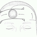
Following admission to the burn center, the patient’s wounds are cleansed with soap and water. Loose tissue and blisters are debrided. Body and facial hair are shaved if involved in the area of a burn. Daily wound care is performed on a shower table with soap and tap water or, if the burn wound is small, at the patient’s bedside following a shower. The use of tanks for wound care has fallen out of favor because of the risks of cross-contamination.

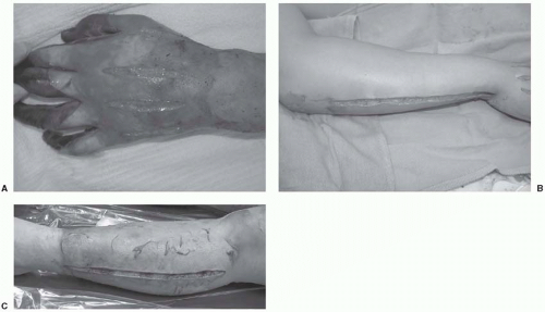
Burn injury destroys the body’s protective layer from the environment and dressings are needed to protect the body from infection and minimize evaporative heat loss from the body. The ideal dressing if it existed would be inexpensive, easy to use, require infrequent changes, and be comfortable. While a number of topical agents are available for burn wound care, it is best to have a simple, well-reasoned wound care plan.

The choice of topical burn wound treatment is contingent on the depth of burn injury and the goals of management. Superficial burn wounds (such as sunburns) require soothing lotions that will expedite epithelial repair such as aloe vera. Partial thickness burn wounds need coverage with agents that will keep the wound moist and provide antimicrobial protection. Deeper partial thickness burn wounds should be covered with agents that will protect the eschar from microbial colonization. Once the eschar has lifted and the wound has begun to epithelialize, a dressing that optimizes epithelialization (i.e., greasy gauze and antibiotic ointment) is utilized. Full thickness burns are also covered with a topical agent that protects the burn wound from getting infected until the time of burn excision.

Prophylactic systemic antibiotics have no role in the management of burn wounds. In fact, the use of prophylactic
antibiotics has been shown to increase the risk of opportunistic infection.1 Since burn eschar has no microcirculation, there is no mechanism for the delivery of systemically administered antibiotics. Therefore, topical agents need to provide broad-spectrum antimicrobial coverage at the site of colonization–the eschar.


FIGURE 15.5. The location of escharotomy incisions on the (A) upper extremity; (B) hand; and (C) lower extremity.

In the early postburn period, the dominant colonizing organisms are staphylococci and streptococci–typical skin flora. Over time, however, the burn wound becomes colonized with gram-negative organisms. Thus, topical antimicrobial agents used in early burn care should have broad-spectrum coverage to minimize colonization of the wound, but they need not penetrate the burn eschar deeply.

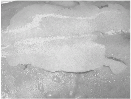
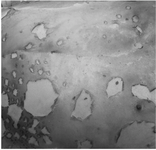
Silver sulfadiazine is the most commonly used topical antimicrobial agent. Silver sulfadiazine has broad-spectrum antimicrobial coverage, with excellent Staphylococcus and Streptococcus coverage. However, silver sulfadiazine is incapable of eschar penetration, so it is less useful in the management of the infected burn wound. Wounds treated with silver sulfadiazine will develop a yellowish-gray pseudoeschar that requires removal by cleansing during daily wound care. Traditionally, the principal drawback of silver sulfadiazine was thought to be leukopenia. However, it is not clear whether the leukopenia that occurs results from silver sulfadiazine toxicity or from the margination of leukocytes as part of the body’s systemic inflammatory response to the burn injury. Regardless, the leukopenia is typically self-limited, and therefore, the silver sulfadiazine should not be discontinued. Patients with a documented sulfa allergy may or may not have a reaction to the silver sulfadiazine. If there is concern about an allergy, a small test patch of silver sulfadiazine can be applied. Typically, if there is an allergy, the silver sulfadiazine will be irritating rather than soothing. In addition, a rash could signal a silver sulfadiazine allergy.

Mafenide (Sulfamylon) is another commonly used antimicrobial agent. Mafenide is available as a cream and, more recently, as a 5% solution. Mafenide, like silver sulfadiazine, has a broad antimicrobial spectrum, including gram-positive and gram-negative organisms. In addition, mafenide readily penetrates burn eschar, making it an excellent agent for treating burn wound infections. Mafenide is commonly used on the ears and the nose because of its ability to protect against suppurative chondritis; however, silver sulfadiazine appears to be equally effective in this setting. Since mafenide penetrates eschar well, twice-daily administration is typically necessary. Mafenide-soaked gauze can also be used as a dressing for skin grafts that have been placed over an infected or heavily colonized wound bed. There are two well-recognized drawbacks of mafenide. Mafenide is a potent carbonic anhydrase inhibitor and, therefore, can cause a metabolic acidosis. This problem can confound ventilator management. In addition, the application of mafenide can be painful and therefore its use may be limited in partial thickness burn wounds.

Silver nitrate is another commonly used topical antimicrobial agent. Silver nitrate provides broad-spectrum coverage against gram-positive and gram-negative organisms. It is relatively painless on administration and needs to be applied every 4 hours to keep the dressings moist. Silver nitrate has two principal drawbacks. First, it stains everything it touches black, including linen, floors, walls, and staff’s clothing. Second, since silver nitrate is prepared in water at a relatively hypotonic solution (0.5%), osmolar dilution can occur resulting in hyponatremia and hypochloremia. Therefore, frequent electrolyte monitoring is needed. Rarely, silver nitrate can cause methemoglobinemia. If this occurs silver nitrate should be discontinued.

Bacitracin, neomycin, and polymyxin B ointments are all commonly used for coverage of superficial wounds either alone or with petrolatum gauze to accelerate epithelialization. These ointments are also used routinely in the care of superficial face burns. Mupirocin (Bactroban) is another topical agent that is effective in treating methicillin-resistant Staphylococcus aureus (MRSA). Mupirocin should be used only when there is a culture-proven MRSA infection to avoid the development of resistant infections.


Fluid Resuscitation

Significant burn injury not only results in local tissue injury but also initiates a systemic response that impacts nearly
every organ system. The release of inflammatory mediators (including histamine, prostaglandins, and cytokines) can lead to decreased cardiac output, increased vascular permeability, and alteration of cell membrane potential. The purpose of fluid resuscitation is to provide adequate replacement for fluid lost through the skin and fluid lost into the interstitium from the systemic capillary leak that occurs as part of the body’s inflammatory response. Therefore, significant volumes of intravenous fluid may be required to maintain adequate organ perfusion.

An understanding of burn shock physiology is essential to understanding the rationale for the various formulas that have been described for fluid resuscitation. Burn injury destroys the body’s barrier to evaporative fluid losses and leads to increased cellular permeability in the area of the burn. In addition, in cases of larger burns (>20%), there is systemic response to injury that leads to capillary leakage throughout the body. Arturson2 in 1979 demonstrated that increased capillary permeability occurs both locally and systemically in burns greater than 25%, and Demling3 demonstrated that half of the fluid administered following 50% TBSA burns ends up in uninjured tissue. Therefore, burn resuscitation must account not only for the loss of fluid at the site of injury but also to the leak of fluid throughout the body. These losses are even greater if an inhalation injury is present since there will be increased fluid leak into the lungs as well as an increased release of systemic inflammatory mediators. Capillary leak usually persists through the first 8 to 12 hours following injury.

The use of formal fluid resuscitation is reserved for patients with burns involving more than 15% to 20%. Awake and alert patients with burns less than 20% should be allowed to resuscitate themselves orally as best as possible. A number of approaches using a number of different solutions have been proposed for intravenous fluid resuscitation.


Crystalloid.

The Parkland formula, as described by Baxter, is still the most commonly used method for estimation of fluid requirements (Table 15.4]). The formula (4 cc × weight in kilograms × %TBSA) provides an estimate of fluid required for 24 hours. The fluid administered should be Lactated Ringer’s (LR). LR is relatively hypotonic and contains sodium, potassium, calcium, chloride, and lactate. Sodium chloride is not used because of the risk of inducing a hyperchloremic acidosis. Half the calculated fluid resuscitation should be administered over the first 8 hours and the second half administered over the next 16 hours. Children who weigh less than 15 kg should also receive a maintenance IV rate with dextrose-containing solution since young children do not have adequate glycogen stores.


TABLE 15.4 THE PARKLAND FORMULA FOR FLUID RESUSCITATION


Formula: 4 cc/kg/%TBSA = total fluid to be administered in the first 24 h


½ of fluid should be given in the first 8 h


½ of fluid should be given in the next 16 h


Fluid should be Ringer’s lactate


Sample calculation: 70 kg person with a 50% TBSA burn


4 × 70 × 50 = 14 L of fluid


7 L in the first 8 hours (875 cc/h)


7 L in the next 16 hours (437 cc/h)


• The formula is only a guideline. Fluid administration should be titrated to urine output of 30 cc/h for adults and 1 cc/kg/h for children.


Pediatric patients less than 15 kg should also receive maintenance fluid based on their weight.


It is important to remember that the formula provides merely an estimate of fluid requirements. Fluid should be titrated to achieve a urine output of 30 cc/h in adults and 1 cc/kg/h in children. Therefore, a Foley catheter should be used to accurately track urine output. If urine output is inadequate, the fluid rate should be increased; conversely, if the urine output is greater than 30 cc/h, the fluid rate should be decreased. Fluid boluses should only be used to treat hypotension and should not be used to improve urine output. Patients with deeper, full thickness burns and patients with inhalation injury tend to require higher volumes of resuscitation.

Only gold members can continue reading. Log In or Register to continue

Stay updated, free articles. Join our Telegram channel

Jun 26, 2016 | Posted by in General Surgery | Comments Off on Thermal, Chemical, and Electrical Injuries

Full access? Get Clinical Tree

Get Clinical Tree app for offline access