Flap |
|
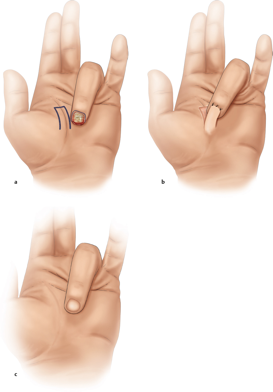
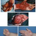
Tissue | Volar skin |
Course of the vessels | Dermal circulation without a named artery |
Dimensions | 1.5 × 1.5 cm |
Extensions and combinations | — |
Anatomy | Relies on the inosculation of the pedicle flap to the wound bed |
Neurovascular pedicle | — |
Artery | — |
Veins | — |
Length and arc of rotation | — |
Diameter | — |
Nerve | — |
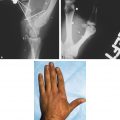
Surgical technique | The flap is elevated at the subcutaneous plane with a pedicle that is no more than twice the length of the base of the flap; it is oriented to inset into the digital pulp defect of the injured finger |
Preoperative examination and markings | — |
Patient position | Supine, with the use of a hand table; tourniquet control |
Dissection | In the subcutaneous plane, with the preservation of perforating vessels (if seen) |
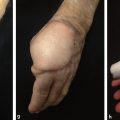
Advantages | Rapid dissection; good color match of skin |
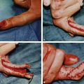
Disadvantages | This is a pedicle flap that requires digital flexion and attachment of the digit to the volar flap for 2–3 weeks |
Pearls and pitfalls | Risk of proximal IP flexion contracture in elderly patients |
Dissection | — |
Extensions and combinations | — |
Contouring and correction | — |
Clinical applications | — |
Plastic Surgery Key
Fastest Plastic Surgery & Dermatology Insight Engine