Flap |
|
Tissue |
Skin or dermal fat flap; can be used as a pedicle (most common) or free flap |
Course of the vessels |
Superficial to the Scarpa fascia, branching in the overlying skin toward the iliac crest |
Dimensions |
10 × 25 cm |
Extensions and combinations |
Usually no combinations with this type of flap; very experienced surgeons may raise the superficial inferior epigastric artery flap as a second skin paddle |
Anatomy |
|
Neurovascular pedicle |
— |
Artery |
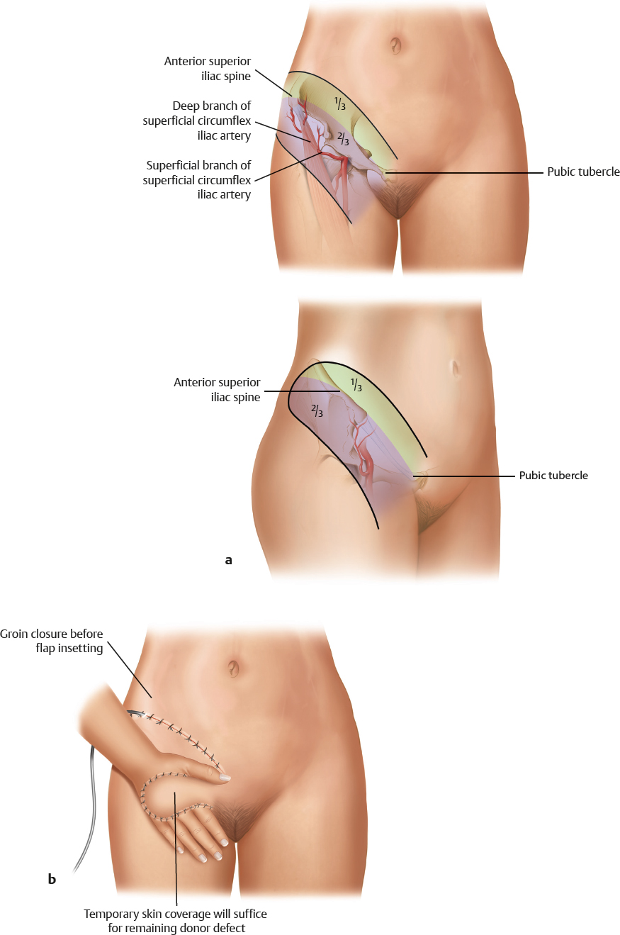
Superficial circumflex iliac artery |
Veins |
Two venous systems: one parallels the superficial circumflex iliac artery and drains into the saphenous bulb, and the other runs deep and directly into the femoral vein |
Length and arc of rotation |
Artery, 1.5–2 cm; veins, 2.5–4 cm |
Diameter |
Artery, 0.8–1.8 mm; veins, 2–3 mm |
Nerve |
Flap is not innervated |
Surgical technique |
|
Preoperative examination and markings |
Create an outline of the flap so that one third is above and two thirds are below the inguinal ligament; the dividing line is drawn from the anterior superior iliac spine to the pubic tubercle |
Patient position |
Supine |
Dissection |
Lateral approach is preferable for a pedicle flap, which is raised from lateral superficial to deep muscle fascia; care must be taken to avoid injury to the pedicle
Medial approach for free flaps: identify the superficial circumflex iliac artery approximately 5 cm below the inguinal line; use a medial incision; identify the superficial vein anterior to Scarpa’s fascia; identify the femoral artery, the superficial inferior epigastric artery, and the superficial circumflex iliac artery; create a lateral skin incision, but leave the deep fascia intact; identify the lateral border of the sartorius muscle; ligate the muscle branches of the deep superficial circumflex iliac artery branch; divide the lateral cutaneous nerve; raise the flap and check for perfusion |
Advantages |
|
Vascular pedicle |
— |
Flap size and shape |
Large flap possible; non–hair-bearing flap |
Combinations |
Medial extensions for hair-bearing flap |
Tissue |
— |
Dissection |
— |
Donor site |
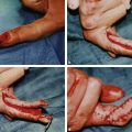
Perfect inconspicuous donor site, with primary wound closure when flap width does not exceed 10 cm |
Further options |
— |
Disadvantages |
|
Bulkiness |
Medial bulk |
Donor site morbidity |
Anesthesia in the lateral cutaneous nerve distribution area |
Flap |
Poor color match in exposed areas |
Pedicle |
Very short pedicle with variable arterial anatomy; arterial diameter is small, and vein grafts are frequently required |
Pearls and pitfalls |
|
Dissection |
Identification of pedicle should precede flap harvest when used as a free flap |
Extensions and combinations |
— |
Contouring and correction |
Correction and debulking are frequently indicated; color match |
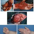
Clinical applications |
Pedicle flap: dorsal hand and forearm defects in younger patients; free flap: dorsal hand and forearm defects in older patients when a short pedicle is possible; not recommended as a pedicle flap in older patients (risk of shoulder stiffness) |