Flap |
|
Tissue |
Muscle or fascia (lower three muscle slips) |
Course of the vessels |
On the muscle surface |
Dimensions |
10 × 15 cm (muscle flap); 10 × 18 cm (fascial flap) |
Extensions and combinations |
Skin island; vascularized ribs |
Anatomy |
|
Neurovascular pedicle |
– |
Artery |
Serratus arcade as extension of the thoracodorsal pedicle; direct serratus branches of the thoracodorsal artery in > 97% of patients |
Veins |
Venae comitantes |
Length and arc of rotation |
≤ 16 cm (when a thoracodorsal pedicle is harvested) |
Diameter |
When thoracodorsal pedicle is harvested: artery, 3.5.4.5 mm; vein: 4.6 mm
When only serratus arcade is taken: artery, 1.1.5 mm; vein, 1.1.5 mm |
Nerve |
Long thoracic nerve (does not always have to be included in the flap) |
Surgical technique |
|
Preoperative examination and markings |
Mark the anterior border of the latissimus dorsi muscle at the tip of the scapula and the 5th through 8th ribs |
Patient position |
Lateral, with the arm elevated at 90 degrees |
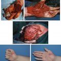
Dissection |
Muscle flap: make a slightly curved incision along the border of the latissimus muscle; identify the muscle border and the serratus arcade; check if the thoracodorsal pedicle is intact; determine the entrance points of motor fibers into the muscle; outline the flap size on the muscle surface; make a medial incision into the muscle; use ligation, coagulation, or clipping of the intercostal vessels to minimize bleeding; release the muscle from the thoracic wall; preserve three proximal slips to prevent wing scapula; dissect the thoracodorsal pedicle to the length required; check the flap for perfusion; transfer the flap
Fascial flap: make a slightly curved incision along the border of the latissimus muscle; identify the muscle border and the serratus arcade; check if the thoracodorsal pedicle is intact; determine the entrance points of motor fibers into the muscle; outline the flap size on the muscle surface; raise the fascia from the muscle surface; coagulate the smaller vessels; preserve the motor nerve; dissect the thoracodorsal pedicle to the required length; check the flap for perfusion; transfer the flap |
Advantages |
|
Vascular pedicle |
Very long pedicle possible; extremely reliable |
Flap size and shape |
Thin and pliable as a fascial flap; minimal donor morbidity |
Combinations |
Vascularized ribs can be harvested with the flap; a small skin island can be included as a monitor island; any combination with other flaps from the subscapular system is possible |
Disadvantages |
|
Flap |
Dissection can be tedious due to many small intercostal connections; injury to the motor nerve may cause wing scapula; the fascia is delicate and can easily be perforated |
Bulkiness |
The muscle flap can be bulky |
Donor site morbidity |
Acceptable; no functional loss except when wing scapula occurs; donor scar is inconspicuous |
Pearls and pitfalls |
|
Dissection |
Identify where the motor fiber enters the muscle; avoid injury to the nerve; the nerve runs laterally from the vascular pedicle; preserve the upper muscle slips; the flaps tend to bleed profusely as fascial flaps; delayed secondary skin grafting is recommended |
Extensions and combinations |
Bone defects can be simultaneously reconstructed with vascularized rib grafts |
Contouring and correction |
Rarely required |
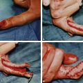
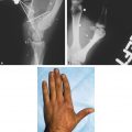
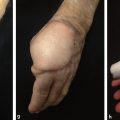
Clinical applications |
Perfect for mid-sized defects that require thin and pliable tissue; gliding tissue for tendon reconstruction; a fascial flap that is mechanically stable can be used for defects of the dorsum of the hand and forearm as well as exposed elbow joints |