Chapter 50 Table 50.1 Temporal fascial flap
Temporal Fascial Flap
Flap |
|
Tissue | Fascia (thickness, 1.5–3 mm) |
Course of the vessels | Subcutaneously on the fascia, from preauricular into the temporal fossa |
Dimensions | 8 × 15 cm |
Extensions and combinations | Can be combined with the deep fascial layer or calvarial bone |
Anatomy |
|
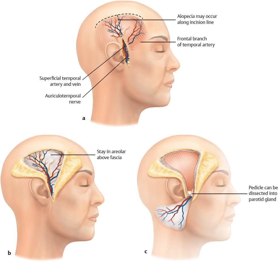
Neurovascular pedicle | Common pedicle with deep fascia (proximal branch of superficial temporal vein/superficial temporal artery); there are no communicating vessels distal to the common pedicle |
Artery | Superficial temporal artery (terminal branch of the carotid artery) |
Veins | Superficial temporal vein |
Length and arc of rotation | 2–4 cm without incising the parotid gland |
Diameter | Artery, 1.5–2.7 mm; vein, 2.0–3.2 mm |
Nerve | Auriculotemporal nerve is included in the fascial layer, but the flap is not innervated |
Surgical technique |
|
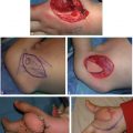
Preoperative examination and markings | Doppler identification of the course of the vessels; marking of the incision line parallel to the hair follicles; outline of flap dimensions |
Patient position | Supine, with the head slightly tilted to the opposite side |
Dissection | Use a T-shaped outline; start the incision by raising a pretragal skin flap; identify and spare the superficial temporal vein anterior and exterior to the superficial temporal artery; identify the superficial temporal artery; proceed with the dissection cephalad, deep to the hair follicles; avoid damage to the very superficial vein; use bipolar coagulation for terminal branches to subdermal plexus; do not damage the frontal branch of the facial nerve; after the cephalad completion of the dissection, incise the flap; lift the flap from the deep fascial plane toward the auricle; observe the flap for perfusion after the completed dissection |
Advantages |
|
Vascular pedicle | Reliable pedicle with sufficient caliber and length |
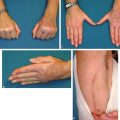
Flap size and shape | Considerable flap size that can cover, for example, the entire dorsum of the hand without bulk; dissection of the flap and donor site can be carried out simultaneously |
Combinations | Can be combined with the deep temporal fascial layer: in this case, the middle temporal vessel at the level of the zygoma has to be preserved; possible combination with calvarial bone graft |
Tissue | Flap is thin and pliable; cover without bulk |
Donor site | |
Disadvantages |
|
Flap size | — |
Donor site morbidity | Frontal nerve may be damaged during dissection; alopecia may result if the superficial plane of the dissection is too close to the hair follicles |
Dissection | — |
Flap | Capillary bleeding may jeopardize graft take |
Pedicle | Pedicle is short; vein is easy to damage due to it superficial location; sometimes vein is absent |
Pearls and pitfalls |
|
Dissection | Watch out for the superficial temporal vein |
Extensions and combinations | When combined with the deep layer, preserve the middle temporal vessel |
Contouring and correction | Contour correction almost never indicated; delayed skin grafting recommended due to tendency for edema and capillary bleeding |
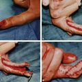
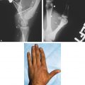
Clinical applications | Dorsum of the hand; deep defects of the palm; degloving injuries of the digits; gliding tissue in scarred wound beds |