The Urinary Tract in Pregnancy

37 The Urinary Tract in Pregnancy


ANATOMY OF THE URINARY TRACT IN PREGNANCY


The kidneys and urinary tract play a major role in maternal adaptation to pregnancy. Consequently, the clinician must understand that observed differences in function cannot be judged by nonpregnant standards. The renal system increases in size and capacity during pregnancy. Anatomic changes involving the urinary tract begin in the first trimester of pregnancy and can persist up to 16 weeks’ postpartum. Intravenous pyelograms (IVPs) performed immediately postpartum demonstrate a 1- to 1.5-cm increase in renal length regardless of the size of the individual. Autopsy studies report an average kidney weight of 307 g in pregnant women, compared with 259 g for kidneys in nonpregnant women. The increase in functional demand (a 50% increase in glomerular filtration rate [GFR]) stimulates renal cell hyperplasia and an increase in proximal tube length much like the renal growth that occurs after a unilateral nephrectomy. In addition, increased water content explains a portion of the increase in the size and weight of the kidney.


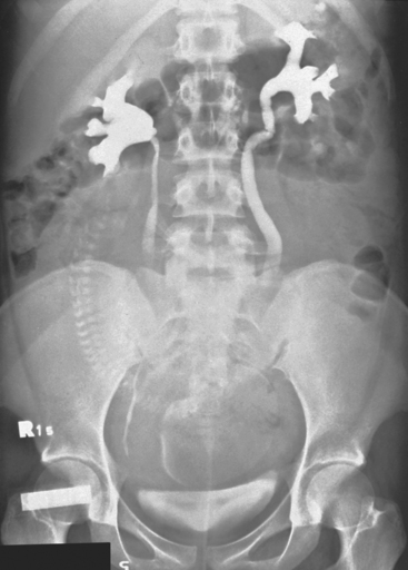
The most striking anatomic change in the urinary tract is dilation of the ureters (Fig. 37-1). Bilateral dilation of the calyces, renal pelvis, and ureters can be seen early in the first trimester and is present in 90% of women in the late third trimester or early puerperium. The changes are usually more prominent on the right and may persist for 3 to 4 months. In 11% of women, ureteral dilation persists indefinitely. In addition, there is reduced ureteral peristalsis and a greater volume of residual urine compared with the nonpregnant state. It is not known whether these patients suffer adverse sequelae, such as persistent asymptomatic bacteriuria from persistent ureteral dilation.


Vesicoureteric reflux is a sporadic, transient occurrence during pregnancy and has been demonstrated radiologically in 7 of 200 (3.5%) pregnant women; the authors felt that this incidence was an underestimate. The enlarging uterus displaces the ureters laterally, and the intravesical ureters are shortened and enter the bladder perpendicularly rather than obliquely. Consequently, the ureterovesical junction is less efficient in preventing reflux. This increased incidence of reflux may explain the high incidence of pyelonephritis during pregnancy; however, this association has not been confirmed. The transitory nature of vesicoureteral reflux and the necessary exposure to x-rays for study purposes hinders adequate evaluation of the problem. Nevertheless, vesicoureteric reflux probably plays only a small role in symptomatic or asymptomatic urinary tract infection.


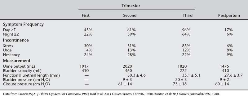
The capacity of the urinary tract increases during pregnancy. Bladder volume during pregnancy increases to 450 to 650 mL, compared with 400 mL in nonpregnant controls (Table 37-1). The hydronephrotic ureters can hold as much as 200 mL extra urine; however, no changes appear in the contraction patterns on retrograde cystometry. Depending on maternal position, uterine size, and position of the fetus, the functional volume of the bladder and ureters is dynamic in the third trimester. This increased functional volume, coupled with high urine flows (especially with fluid mobilization at night), causes polyuria and nocturia in most pregnant women.


The etiology of ureteral and bladder dilation generates much discussion. Sharp termination of the ureteral dilation at the pelvic brim seen on IVP suggests an obstruction. When a woman is upright or supine, as during the filming of an IVP, the pregnant uterus compresses the ureter against the pelvic rim and its overlying iliac vessels. On the left side, the ureter is somewhat protected by the iliac arteries and sigmoid colon and, as a result, is usually less dilated than the right ureter. Although mechanical obstruction plays a major role in ureteral dilation during pregnancy, the relative infrequency of ureteral obstruction by large ovarian tumors or fibroids in nonpregnant women suggests additional factors. In addition, high urine production, as occurs in diabetes insipidus or pregnancy, is also associated with urinary tract dilation.


In the past, the elevated progesterone levels that accompany pregnancy were thought to cause smooth muscle relaxation and subsequent hypotonicity and hypomotility of the ureter, defects that would contribute to ureteral dilation. Contrary to the latter observation, the large doses of synthetic progesterone used in cancer chemotherapy do not cause ureteral dilation. Measurements of ureteral tone during pregnancy reveal an increase in ureteral tone and no decrease in frequency or amplitude of ureteral contractions. Histologic study of the ureters of pregnant animals reveals smooth muscle hypertrophy and hyperplasia of the connective tissue. Thus, progesterone probably plays a small role in ureteric dilation during pregnancy.


RENAL CHANGES IN PREGNANCY


Much of the data relating to the effect of pregnancy on renal hemodynamics are derived from small studies in which measurements show considerable individual variation. Autoregulation maintains renal blood flow at a relatively constant level despite wide variations in perfusion pressure (mean renal artery pressure). Renal blood flow is usually assessed by ρ-aminohippurate clearance, which measures effective renal plasma flow (ERPF). The ERPF significantly increases during pregnancy. It reaches a peak increment in midtrimester of 50% to 85% and then shows a small decline during the third trimester that is unrelated to posture. The ERPF and GFR in pregnancy are markedly affected by posture, being maximal when the pregnant woman lies on her side. Normal pregnancy is associated with plasma volume expansion and an increase in the GFR of 40% to 65% (measured by insulin clearance) and a decrease in GFR of approximately 15% to 20% late in the third trimester. The mechanisms responsible for the increase in GFR, plasma volume, and renal plasma flow rate are unknown. Nitric oxide, endothelin, and relaxin may play a role in renal vasodilation in human pregnancy. Changes in renal anatomy, hemodynamics, and tubular function are listed in Box 37-1.


BOX 37-1 PHYSIOLOGIC CHANGES IN PREGNANCY


TESTS OF RENAL FUNCTION IN PREGNANCY


Tests of renal function in pregnancy must be interpreted in relation to the changes in plasma volume, glomerular filtration, and tubular reabsorption that normally occur with advancing gestation. Many of the commonly used tests of function yield lower results in pregnancy than in the nonpregnant state. Consequently, values that may be regarded as normal in the nonpregnant state may well indicate renal dysfunction in pregnancy.


Uric acid, blood urea nitrogen (BUN), and serum creatinine levels are crude indices of renal function. In pregnancy, plasma uric acid usually decreases by 25% beginning in the first trimester and increases during the third trimester. Upper normal limits of plasma uric acid levels are 5 to 5.5 mg/dL in pregnancy. Levels are influenced by race, multiple gestation, and time of day sampled, with higher levels in the morning. An indicator of renal filtration, the BUN normally decreases from nonpregnant levels of 12 mg/dL (4.3 mmol/L) to 9 mg/dL (3.2 mmol/L), and plasma creatinine levels decline from a nonpregnant mean value of 0.7 mg/dL (62 mmol/L) to 0.5 mg/dL (44 mmol/L). If the plasma creatinine level exceeds 0.9 mg/dL or if the BUN is greater than 14 mg/dL at any stage in pregnancy, renal dysfunction should be suspected and more detailed investigation should be performed.


The 24-hour creatinine clearance is the best clinical measurement of GFR. By week 8 of pregnancy, the creatinine clearance rate normally increases by 45% and remains elevated during the second trimester. In the final weeks of pregnancy, creatinine clearance usually declines to near nonpregnant levels (Fig. 37-2).
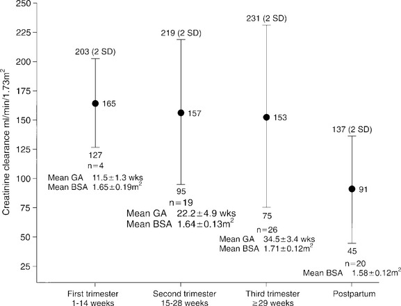


Error in the measurement of creatinine clearance in a pregnant woman can occur; the most common type of error is an incomplete 24-hour urine collection. Accurate timed urine collection is particularly difficult in pregnancy because significant volumes of urine may remain in the dilated collecting system. To avoid this error, patients should be well hydrated and should rest on their left side for 1 hour before starting and completing the 24-hour urine collection. In addition, the secretory component of creatinine excretion increases in moderate renal failure, and creatinine clearance rates tend to overestimate GFR. Despite these problems, creatinine clearance still remains the most useful measure of GFR in clinical practice.


Urinalysis is essentially unchanged during pregnancy. However, many variables can affect the results. Normal kidneys should be able to concentrate urine to a specific gravity of 1.026 or more and to dilute urine to a value less than 1.005. In pregnancy, posture affects urine concentration and specific gravity. Urine tends to be more dilute after a left lateral position is maintained compared with an upright position. The urine must be at room temperature for the dipsticks to be reliable. Dipsticks exposed to air will give false-positive results for glucose and false-negative results for blood. Observational error and training also affect the sensitivity of predicting proteinuria by dipstick. Saudan et al. (1997) showed that the use of an automated urinalysis device for the detection of proteinuria reduced the false-positive rate. Proteinuria diagnosed on dipstick should be confirmed with a 24-hour urine collection.


The method of collection is very important when collecting a urine specimen. It is difficult for the woman to obtain a satisfactory clean voided specimen by herself, especially when she is far along in pregnancy. In addition, the specimen must be collected before the pelvic examination; it may be collected by the examiner while the patient is in the dorsal lithotomy position.


Laboratory investigation begins with urinalysis of protein, glucose, ketones, specific gravity, and sediment. Glucosuria is usually detected with a glucose oxidase–impregnated dipstick. In pregnancy, the most common reasons for persistent glucose in the urine are physiologic glucosuria of pregnancy and diabetes. However, the possibility of primary renal disease with renal glucosuria should be considered. Conventional screening for proteinuria uses a dipstick that is sensitive to albumin. Five percent of healthy adults exhibit postural proteinuria, a benign condition; this can be ruled out by comparing protein levels in the first voided urine with a specimen obtained after the woman was upright for several hours. False-positive results for protein can be due to concentrated urine, many white blood cells in the urine, or vaginal secretions with epithelial cells. Fever, stress, and exercise can also cause transient proteinuria.


Proteinuria in pregnancy should be evaluated using a 24-hour urine collection and should not be considered pathologic until it exceeds 300 to 500 mg in 24 hours. Higby et al. (1994) showed that the upper limit or normal was 260 mg for urinary protein and 29 mg for albumin in a 24-hour period. Both increased after 20 weeks’ gestation. Studies have shown that a 2-hour or 12-hour collection of urine correlates with creatinine clearance and protein measured in a 24-hour specimen. Evans and associates calculated total protein using a protein/creatinine ratio in the 2-hour group and compared the results with the 24-hour urine (1840.8 ± 786 and 1944 ± 1060 mg [mean ± SE], respectively, r2 = 0.95, p < .0001). The nephritic syndrome is characterized by greater than 3 to 3.5 g/24 hours. In assessing the significance of proteinuria in pregnancy, the clinician should remember that increasing protein excretion with advancing gestation associated with known renal disease does not necessarily indicate significant progression of the disease.


URINARY TRACT DISEASES IN PREGNANCY


Diseases of the urinary tract and kidneys constitute a major portion of obstetric complications and may be classified as infection, renal disease, and urologic disease.


Urinary Tract Infection


PATHOPHYSIOLOGY


In order to appreciate the frequency and clinical significance of urinary tract infections (UTIs) during pregnancy, it is important to review such findings in nonpregnant women. During any given year, 11% of all women report having had a UTI and, during their lifetime, at least 50% of women will suffer one or more UTI. The prevalence of asymptomatic bacteriuria in young women is about 4% to 6%. It is usually associated with the same risk factors as for symptomatic UTI; these include the use of diaphragm plus spermicide and sexual intercourse. Approximately 8% of women with asymptomatic bacteriuria develop a symptomatic infection within 1 week of detection. Escherichia coli is the most common pathogen (80% of cases) in these women. The rate of symptomatic infection increases to 15% if there is associated pyuria.


After an initial urinary tract infection, about 25% to t50% of women will have another infection within 1 year. Acute cystitis is the most common UTI, which is characterized by dysuria, urinary frequency, and hematuria. The most common risk factors for acute cystitis are a previous history of cystitis and frequent of recent sexual intercourse.


Acute pyelonephritis is a common complication in women and is responsible for a large number of office consultations with physicians and a large number of hospital admissions every year in the United States. In general, most nonpregnant women with pyelonephritis are treated in ambulatory settings. Risk factors for pyelonephritis in these women include a family history of UTI, diabetes mellitus, and the presence of urinary incontinence.


The physiologic changes of pregnancy increase the likelihood of symptomatic upper urinary tract disease, resulting in maternal and fetal morbidity and, occasionally, mortality. In fact, UTIs are one of the most common medical complications of pregnancy (Table 37-2). Thus, an understanding of the pathogenesis, clinical presentation, diagnosis, therapy, and prognosis is essential.


Table 37-2 Incidence of Urinary Tract Infection During Pregnancy


Infection Incidence (%)
Asymptomatic bacteriuria 2–11
Acute cystitis 1–4
Acute pyelonephritis 1–2

Although bloodborne organisms (e.g., staphylococcal bacteremia) may occasionally infect the renal parenchyma, the most common route of infection is ascension up the urinary tract. Female anatomy and behavior set the stage for inoculation of the bladder. The urethra (3-4 cm in length) is close to the vagina and rectum; both are fertile reservoirs for uropathogens. Indeed, the presence of Enterobacteriaceae at the vaginal vestibule is a predictor of asymptomatic bacteriuria. Pumping action by intravaginal coitus or urethral massage allows inoculation of the bladder.


Inoculation of the bladder does not always lead to colonization or symptomatic disease. Host-parasite interactions determine the likelihood of infection. The presence or absence of bacterial virulence factors may explain why some women with asymptomatic urinary tract infection go on to develop symptoms. These factors have been best defined for E. coli, the most common uropathogen, and include increased adherence to uroepithelial cells, high quantities of K-antigen, the presence of aerobactin, and hemolysin production. Of these, adhesive properties seem to be the most important. E. coli pyelonephritis isolates adhere to uroepithelial cells better than do E. coli cystitis isolates, and urinary isolates tend to adhere better than do random fecal E. coli isolates. This adhesive capacity is mediated by the presence of adhesions on the bacterial cell surface. Often these adhesions are pili or fimbriae. Pili or fimbriae bind to the β-globoseries glycolipid receptors on the surface of the uroepithelial cells.


In nonpregnant women, 10% to 20% of E. coli strains isolated from patients with cystitis or asymptomatic bacteriuria express P-fimbriae. On the other hand, 80% to 90% of strains isolated from acute, nonobstructive, pyelonephritis express P-fimbriae. Recently, the same pattern has been shown in pregnant women.


Usually, the normal urinary tract resists colonization by bacteria and rapidly and efficiently eliminates microorganisms that gain access to the bladder. Urine has antibacterial activity, extremes in osmolarity, high urea concentrations, and low pH levels that inhibit the growth of uropathogens. However, pregnancy makes the urine more suitable for bacterial growth by increasing pH and normalizing osmolarity.


DIAGNOSIS


The diagnosis of urinary tract infection has been based on the landmark studies by Kass (1956) and Elder et al. (1971) at Boston City Hospital. In a population of asymptomatic pregnant and nonpregnant women, a bacterial colony count of ≥105 colony-forming units (cfu) per milliliter in two or more clean-catch midstream urine (MSU) specimens reliably distinguished infection from contamination. Between 10% and 20% of pregnant women with an initial positive culture have a second negative culture within a week, even without antibiotic therapy. Furthermore, 95% of patients with clinical pyelonephritis have persistent positive cultures at ≥105 cfu/mL. Since these studies, most clinicians have used the criterion of 105 cfu/mL on a clean-catch MSU specimen to diagnose UTI. However, this criterion has not proved to be sufficiently predictive in nonpregnant women with acute dysuria or infections with fastidious organisms, or in catheterized patients.


Suprapubic or urethral aspiration of urine has been used to prevent vaginal contamination. In a series of classic articles, Stamm et al. (1982, 1989) demonstrated the following in acutely dysuric nonpregnant women: the correlation between MSU colony counts and bladder bacteria (by suprapubic or urethral aspiration) was 0.78; in women who were dysuric and whose bladders contained coliform bacteria, approximately 50% have MSU <105 cfu/mL; and an MSU with ≥102 cfu/mL predicted most accurately a positive culture by bladder aspiration (sensitivity 0.95 and specificity 0.85). In contrast, an MSU with ≥105 cfu/mL had a sensitivity of 0.51 and a specificity of 0.99; pyuria (≥8 leukocytes/mm3) on an unspun MSU specimen was highly sensitive (0.91) but not specific (0.5); over half of women have an MSU culture positive for more than one organism; and 48% of patients with more than one organism isolated on MSU, “a contaminated urine,” have coliform ≥102 cfu/mL on bladder or urethral aspiration.


With the criterion of ≥105 cfu/mL, the urine culture cannot be viewed as a precise quantitative assay. The lack of precision results from several causes, including the type or stage of the disease (e.g., asymptomatic bacteriuria vs clinical pyelonephritis); obstructions or abnormalities of the urinary tract; perinephric abscess; urolithiasis; acidification of the urine; hydration and diuresis; polyuria; collection methods (e.g., MSU vs suprapubic aspiration); transport, storage, or culture methods; and fastidious organisms.


Asymptomatic bacteriuria with aerobic organisms has been associated with adverse pregnancy outcome. Many of the preceding fastidious organisms in the vagina or cervix are also associated with adverse pregnancy outcome and, at least with group B streptococcus, a UTI is more often associated with adverse pregnancy outcome than is vaginal colonization alone. It is tempting to speculate that UTIs with fastidious organisms may also predict adverse pregnancy outcome, but neither the correlation between vaginal and urinary tract colonization nor the correlation between fastidious organisms in the urinary tract and adverse pregnancy outcome has been studied.


The classic criterion of ≥105 cfu/mL for diagnosing UTI is challenged in catheterized women. Stark and Maki (1984) demonstrated that 96% of patients with low levels of coliform bacteriuria (<105 cfu/mL) progressed to ≥105 cfu/mL within 3 days if they did not receive antibiotics and remained catheterized. Although these observations help with the interpretation and management of positive urine cultures in catheterized patients, the questions of who and when to sample the urine in catheterized patients remain unanswered.


Urine cultures delay definitive diagnosis, are expensive ($50–$70), and require microbiologic technology. Thus, culture-independent diagnostic tools have appeal. The most commonly used tests include microscopic examination of urine, measurement of leukocyte esterase and nitrite, filter isolation of bacteria and white blood cells, and screening for bioluminescence.


For years, the technique for rapid diagnosis has been microscopic examination of the urine. A Gram stain is performed by placing a drop of uncentrifuged urine on a slide. A positive Gram stain is ≥3 bacteria per oil immersion field (1000 × magnification). The sensitivity (84%–94%) and specificity (68%–97%) of this technique in predicting a culture positive at 105 cfu/mL compare favorably with those of other newer tests. In addition, the Gram stain and examination of unstrained urinary sediment after centrifugation can identify the most probable pathogen (e.g., gram-negative rods) and the presence of upper urinary tract disease (e.g., white cell casts). The test takes about 5 minutes to perform and costs very little ($20).


The least expensive and least labor-intensive of the other rapid tests is the test for nitrite and leukocyte esterase, Chemstrip LN (Biodynamics; Indianapolis, IN). The plastic dipstick contains patches of color-responsive reagents that identify esterase and nitrite. The test takes 2 minutes to perform and costs approximately $25 per test. At a threshold of ≥105 cfu/mL, the leukocyte esterase-nitrite strip has a sensitivity of 60% to 100% and a specificity of 60% to 98%. At a threshold of ≥103 cfu/mL, the strip has a sensitivity of 52% to 73% and specificity of 68% to 83%.


The filter isolation test, Bac-T-Screen (Marion Laboratories, Inc., Kansas City, MO), uses an instrument ($2000) to filter 1 mL of urine. The attached bacteria and sediment (white blood cells) are stained and decolorized. The residual stain is proportional to the amount of bacteria and number of white blood cells in the urine. The test can be performed in 2 to 3 minutes and costs $85 per test. At a culture threshold of ≥105 cfu/mL, the Bac-T-Screen has a sensitivity of 85% to 96% and a specificity of 38% to 81%. At a culture threshold of ≥103 cfu/mL, the sensitivity is 74% and specificity is 78%.


The bioluminescence tests (e.g., 3M LUMAC [Biocounter, 3M Company, St Paul, MN]) are based on the principle that bacteria and mammals have distinct adenosine triphosphate (ATP) that can be destroyed selectively. After destruction of mammalian ATP, bacterial ATP can be detected by the bioluminescence produced in the firefly luciferin-luciferase reaction. The test takes about 30 minutes to produce a result, and the instrument costs $9000. The price of the reagents is approximately $1.60 per test. At a culture threshold of ≥105 cfu/mL, the test has a sensitivity of 93% to 99% and a specificity of 81% to 96%. At a culture threshold of ≥104 cfu/mL, the test has a sensitivity of 88% to 95% and a specificity of 81% to 95%.


The essential question is whether culture-independent tests are sufficiently robust to replace or enhance urine culture in the diagnosis of urinary tract infection. Table 37-3 depicts the efficiency of culture-independent tests at common prevalences of urinary infection at ≥105 cfu/mL: 5% to reflect the prevalence of asymptomatic bacteriuria in pregnancy and 50% to reflect the prevalence of positive culture in women with dysuria. A false-negative rate of 6% to 20% with the culture-independent tests does not qualify them to supplant urine culture; however, many clinicians recommend that acutely dysuric nonpregnant women be treated without a culture. In pregnancy, the acutely dysuric women should always have a culture because 10% to 15% of positive cultures have group B streptococcus, an organism that predicts adverse pregnancy outcome.


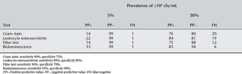
At a prevalence of 2% to 10%, as is seen in asymptomatic bacteriuria during pregnancy, the positive predictive values of culture-independent tests drop precipitously in both theory (see Table 37-3) and practice and should not be used for diagnosis. On the other hand, the negative predictive value is 98% or more with any of these tests. In a low-risk population, urine testing for leukocyte esterase and nitrite on a clean-catch, first-void midstream specimen can supplant urine culture. In high-risk groups (Box 37-2), a culture should be obtained each trimester.


ASYMPTOMATIC BACTERIURIA


A combination of host defense inefficiency, anatomy, behavior, and microbial virulence factors identifies a cohort of women who will have episodes of bacteriuria throughout their lifetimes. Cross-sectional prevalence studies identify 1% to 8% of women with asymptomatic bacteriuria. In longitudinal studies, 30% to 50% of nonpregnant women with bacteriuria have symptomatic lower tract infections during 3 to 5 years of follow-up. Most episodes cluster over a 3- to 4-month period followed by an asymptomatic interval of variable length. Studies with 9- to 19-year follow-up on 60 asymptomatic bacteriuric schoolgirls (6 to 10 years old) were compared with studies on 38 nonbacteriuric control schoolgirls matched for age, race, and school. Episodes of bacteriuria in the 5-year study period for infected girls and controls were five or more episodes (22% and 3%, respectively), and episodes during pregnancy (64% and 27%, respectively). Interestingly, the children of bacteriuric women were more likely to have UTIs than were the children of controls.


Between 20% and 30% of women who are bacteriuric during pregnancy will be bacteriuric on long-term follow-up cultures when not pregnant. Radiologic examination at follow-up of women who were bacteriuric during pregnancy revealed abnormalities in 316 (41%) of 777 women (range, 5%–75%). Chronic pyelonephritis was the most common radiologic diagnosis (47% of abnormalities). The incidence of bacteriuria during first pregnancies was significantly greater in women with (47%) than without (27%) renal scarring from childhood urinary infections. Similar controls who had not had childhood urinary infections had an incidence of 2%.


The cohort of women with chronic, episodic bacteriuria is identified by routine screening of urine cultures at the first prenatal visit. The prevalence of asymptomatic bacteriuria (two or more cultures at ≥105 cfu/mL) is increased by prior renal or urinary tract disease, diabetes, sickle cell trait or disease, poor hygiene, high parity, increased age, and lower socioeconomic status. The overall prevalence varies between 1.9% and 11.8%, with the lowest prevalence in primiparous patients of the upper socioeconomic class and the highest among indigent multiparas. Although most women with asymptomatic bacteriuria are identified shortly after entering prenatal care, approximately 1% to 2% acquire bacteriuria later in pregnancy.


The microbiology of urinary tract infections in pregnancy is summarized in Table 37-4. The predominant organism is E. coli, and the identification markers and virulence traits from strains isolated from pregnant women with pyelonephritis do not differ significantly from those found in strains isolated from nonpregnant women with pyelonephritis. The pyelonephritic E. coli strains and strains from asymptomatic bacteriuria or cystitis patients differed in resistance to serum antibodies (83% vs 51%, P < .05) and epithelial adherence (63% vs 19%, P < .001).


Table 37-4 Microbiology of Urinary Tract Infections in Pregnancy


Organism Percentage
Escherichia coli 60–80
Klebsiella pneumoniae-Enterobacter 3–5
Proteus sp. 1–5
Streptococcus faecalis 1–4
Group B streptococcus 4–8
Staphylococcus saprophyticus 1–3

The isolation and concentration of organisms other than gram-negative rods depend on preparation (cleansing the urethral orifice), collection methods (midstream vs suprapubic aspiration), and selective medium (Todd-Hewitt broth for group B streptococcus). Although E. coli and other gram-negative rods are associated with pyelonephritis during pregnancy, other organisms may be important in other adverse pregnancy outcomes. A large study that uses modern, comprehensive microbiologic techniques is needed to relate specific urinary tract pathogens to pregnancy outcome.


Uncomplicated, asymptomatic bacteriuria is a significant health risk for pregnant but not nonpregnant women. Asymptomatic bacteriuria has been associated with pyelonephritis, preterm birth, hypertension, and fetal neuropathology. The most consistent association is a greater likelihood of pyelonephritis. In 1699 patients with untreated asymptomatic bacteriuria (Sweet [1977

Only gold members can continue reading. Log In or Register to continue

Stay updated, free articles. Join our Telegram channel

Mar 11, 2016 | Posted by in Reconstructive surgery | Comments Off on The Urinary Tract in Pregnancy

Full access? Get Clinical Tree

Get Clinical Tree app for offline access