The Spitz nevus is a relatively common skin lesion in children and is less commonly seen in adults. The lesion is defined by the presence of distinctive-appearing spindle or epithelioid cells on light microscopy in a recognizable nevus-like pattern. Spitz lesions share features with melanoma on light microscopic examination. When Spitz features are atypical or typical features are absent, distinction from melanoma can be difficult. A spectrum of pathology of Spitz lesions can be found from lesions that are benign and typical to lesions that are atypical with melanoma-like features and frank melanoma. There is significant interobserver variation in interpretation of Spitz lesions. The lack of uniformly applied criteria for distinction of light microscopic grades and the confusion in diagnostic terminology demonstrate the difficulty in the pathologic interpretation of these lesions. Exciting progress has been made recently in ancillary testing that will likely be helpful in determining in more detail the biologic nature of these lesions, in better differentiating the benign Spitz lesions from malignant lesions, and in eventually improving treatment recommendations.
The Spitz nevus is a melanocytic lesion that has melanoma-like features on light microscopic examination but in typical form is distinguishable from melanoma, and usually exhibits benign behavior. There are also Spitz-like melanomas with microscopic features of Spitz lesions but otherwise distinctly malignant microscopy. Finally, borderline Spitz lesions have features of Spitz lesions and features of melanoma, and are recognized as of uncertain biologic potential and possible melanoma. These more rare borderline lesions are the most controversial regarding diagnosis and treatment. However, even Spitz nevi with typical histology have on very rare occasions unexpectedly led to metastasis and death. Thus, even though Spitz lesions are easily recognized as distinct, they can defy current light microscopic diagnosis principles that are traditionally used to distinguish those malignant versus nonmalignant. There are many unanswered questions about the biologic nature of Spitz nevi. The diagnosis, prognosis, management, and even nomenclature of these lesions is controversial.
Despite improvements in diagnosis and combined experience over the past century since the Spitz lesion was first described, this area remains one of the most controversial in pathology. To date investigations searching to more accurately diagnose and effectively treat these lesions have included limited numbers of cases and suffer from cohort bias. Multiple diagnostic and management strategies have been proposed and are individually utilized. Recently described investigational techniques seem promising. At this point, multicenter studies are needed to collectively determine whether they can be relied on to more accurately predict prognosis and therefore improve management.
Epidemiology
Spitz nevi are common in children but relatively uncommon in adults. Spitz nevi account for approximately 1% of excised nevi in children. Many different names have been used for Spitz nevi since the original description of juvenile melanoma ( Box 1 ), in an attempt to assign probable behavior. The names reflect a spectrum from benign to malignant, based on light microscopic analysis. Melanoma is probably best reserved in the name for Spitz lesions when referring to frank melanoma that has Spitz features (“spitzoid melanoma”) or in Spitz lesions with features of Spitz and melanoma where biologic behavior is not predictable (“borderline melanoma”). The term “nevus” connotes benign behavior and is probably best reserved for lesions on the benign end of the spectrum or when atypical features are present if used with a qualifying description (“Spitz nevus with atypical features”). The term lesion is more neutral and can more broadly encompass all “Spitz lesions.” Urso and Piepkorn suggest use of the term Spitz “tumor” to distinguish all Spitz lesions as separate entities from nevus and melanoma therby emphasizing separate diagnostic and biologic paths. Individual names are used inconsistently between various investigators, and thus attention should be paid to the individual author’s application of the terminology.
Spitz nevus synonyms
Juvenile melanoma
Spindled and epithelioid cell nevus
Pigmented spindle cell nevus of Reed
Nevus of large spindleoid or epithelioid cells
Synonyms for Spitz that are neutral
Spitz lesion
Spitz tumor
Spitzoid lesion; spitzoid tumor; spitzoid melanocytoma; spitzoid neoplasm
Atypical or borderline Spitz synonyms
Atypical Spitz nevus
Melanocytic tumor of uncertain potential (MELTUMP); melanocytic lesion of uncertain malignant potential (MUMP); Spitz tumor if uncertain potential (STUMP)
Borderline melanoma
History
The Spitz nevus has been recognized in the literature since the early twentieth century, when Darier and Civatte described a fast-growing red nodule on the cheek of a young boy and reported that they were unable to decide by light microscopy whether it was a melanoma or not. The pathology of the lesion they described, which would later be termed a Spitz nevus, was indistinguishable in their viewpoint from melanoma. Similar lesions would normally have been reported as malignant melanoma at this time; however, some researchers such as Darier and Civatte were beginning to recognize that these growths could exhibit benign clinical behavior. Still, the only criterion for distinguishing these lesions from melanoma was age of the patient. In 1948, Sophie Spitz described in detail the light microscopic features of a group of these lesions, which she termed juvenile melanoma, and the diagnosis became widely recognized as distinct. Spitz compared the pathology of juvenile melanoma in individuals 18 months to 12 years old to melanoma in children 14 to 19 years old and to melanocytic nevi in children. She described common features of these juvenile melanoma lesions, and emphasized the presence of giant cells as a distinguishing feature from postpubertal melanoma.
Spitz believed that these juvenile melanomas were unable to metastasize until adulthood because of a hormonal effect. The indicated treatment for juvenile melanoma in these years was removal before adulthood to prevent malignant degeneration. There were 2 problems with this assumption that she would later recognize: (1) a 12-year-old girl included in the original Spitz description died of metastasis, and (2) benign Spitz lesions were found in adults as well. Both these factors implied that age was not the only factor in determining benign verses malignant behavior.
Allen and Spitz later described a refined description of pathologic criteria for distinguishing Spitz nevi from melanoma in 1953. Arthur Allen, Sophie Spitz’s husband, eventually reviewed the pathology of the 12-year-old in the original description who died from melanoma and determined that the pathology was, in fact, not consistent with the diagnosis of juvenile melanoma. Thus, the difficulty in pathologic diagnosis of Spitz nevi was apparent as early as the first description. Although more refined criteria for diagnosis of Spitz nevus are available today, the biologic potential remains unpredictable in a subset of lesions with atypical features that resemble melanoma.
Clinical presentation
Spitz nevi occur most often in children or young adults but can occur at any age. Nearly half to two-thirds of Spitz nevi occur in individuals younger than 20 years. Spitz nevi become less common with increasing age, and they are more likely to be diagnosed as melanoma with increasing age of the patient. Congenital Spitz nevi have been reported. The lesions are more common in Caucasians with fair skin type and are slightly more frequently found in females.
Spitz nevi commonly present on the face, head, neck, or lower extremities, but can occur anywhere on the body. In one study, Spitz nevi in children were more commonly on the head and neck whereas Spitz nevi in adults were more commonly located on the extremities (consistent with the observation in children that melanocytic nevi are found in greater frequency on the head and neck). The lesions are frequently solitary, but multiple and agminated (multiple grouped) lesions can occur. Grouped lesions may coalesce on a base of macular hyperpigmentation. Spitz nevi most often occur de novo but uncommonly can occur in association with an existing melanocytic nevus, usually either a congenital nevus or common melanocytic nevus.
The stereotypical Spitz nevus is a pink papule on the cheek of a child ( Fig. 1 ). Spitz nevi are most commonly pink or flesh-colored owing to their relative paucity of melanin; however, red, brown, black, or brown/black lesions can occur ( Fig. 2 ). The brown/black variant is also termed pigmented spindle cell nevus of Reed, and has been reported as a separate diagnostic entity, although most investigators consider it within the Spitz nevus spectrum.
Spitz nevi are usually flat-topped or dome-shaped papules that are symmetric and well circumscribed. The lesions are usually small (less than 6 mm) but can be larger (1 cm diameter or more). Lesions are generally round, but can be oval or rectangular. Telangiectasia may be associated with the lesion ( Fig. 3 ). The surface can be ulcerated or scabbed ( Fig. 4 ). More unusual clinical appearances such as halo phenomenon, polypoid shape, or pedunculated lesions can occur.
Spitz nevi are generally asymptomatic but growth can occur over a period of months. Eruptive Spitz nevi have also been described. The lesions can uncommonly bleed, itch, or be reported as painful.
Differential diagnosis
The Spitz nevus is commonly misdiagnosed based on clinical impression ( Box 2 ). Red lesions are commonly misdiagnosed as pyogenic granuloma, especially if rapid growth, ulceration, or bleeding is found. Light brown lesions may mimic dermatofibroma and brown/black lesions can be mistaken for atypical nevi. Flesh-colored lesions may be misdiagnosed as a cyst, wart, or amelanotic melanoma. Spitz nevi presenting as a pink papule on the face in young children can be mistaken as a juvenile xanthogranuloma, a common skin lesion in young children. Misdiagnosis of angioma or hemangioma of infancy may also be made; however, clinical distinction should be possible given Spitz nevi have a firmer consistency, are more well demarcated than a hemangioma of infancy, and have a history of onset after the neonatal period.
Pyogenic granuloma (lobular capillary hemangioma)
Hemangioma of infancy
Juvenile xanthogranuloma
Cyst
Wart
Melanoma
Basal cell carcinoma
Melanocytic nevus
Histiocytoma
Metastatic carcinoma
Pseudolymphoma
Primary adnexal tumor
Mastocytoma
Angioma
Diagnosis
The diagnosis of Spitz nevus can be suspected based on clinical presentation, and is confirmed by pathologic examination. As previously mentionid, lessions are, however, frequently misdiagnosed as either pyogenic granuloma or amelanotic melanoma, and the diagnosis is made retrospectively after light microscopic evaluation. If Spitz nevus is suspected, excisional biopsy is preferred in order to examine the entire specimen including the architectural details. Full-thickness removal around the periphery of the lesion and through the dermis down to the mid or deep subcutaneous fat is ideal. Shave or partial excision may obscure pathologic features key to distinction from atypical nevi or melanoma. However, each case should be considered individually for its particular circumstances when biopsy technique is considered to avoid overtreatment, especially in very young children.
Dermoscopic features
Dermoscopy (also known as dermatoscopy, epiluminescence microscopy, and skin surface microscopy) is a form of in vivo microscopy. Although recently described, it is now a routinely utilized diagnostic tool. Skin surface microscopy allows visualization of structures that are not visible with the naked eye. The technique is especially useful for pigmented lesions and amelanotic lesions, and can improve diagnosis or delineate microscopic change in a lesion over time. Terminology used to describe dermoscopic features has been widely described. Several different methods of interpretation of the reflective pattern have been suggested, all combining overall features of symmetry observed within the lesion with detailed diagnostic criteria present or absent in the lesion for a given diagnostic entity. Diagnostic accuracy improves with use of dermoscopy.
The pigmented spindle cell nevus of Reed, a pigmented subset of Spitz nevus, has been well characterized on dermoscopy. Three main patterns have been described: (1) starburst, (2) globular, and (3) atypical. The globular and starburst patterns both consist of a central area surrounded by a prominent regular pigment network on the periphery. The starburst pattern is characterized by regularly spaced pigment globules that appear to stream outward toward the periphery of the lesion. Change from globular to starburst (stellate) pattern has been described over the course of evolution of a lesion, and thus these patterns may represent different stages of the lesion. The central area of pigmentation can have a retiform pattern, or the pattern can be a reverse one characterized by dark holes with light network pattern intervening.
The dermoscopic pattern in nonpigmented Spitz nevi is characterized by the presence of dotted vessels regularly distributed throughout the nevus, and is often associated with network depigmentation. Other nonspecific patterns can be seen as well. The dermoscopic features of nonpigmented Spitz nevi are shared by melanoma, and dermoscopy cannot be used to distinguish these lesions.
Use of dermoscopy can increase the accuracy of diagnosis of Spitz nevus based on globular or starburst subtype pigment patterns. However, Spitz nevi showing an atypical pattern on dermoscopy or a multicomponent pattern are frequently misdiagnosed as other melanocytic nevi, skin neoplasms, or even nonmelanocytic lesions such as inflammatory conditions. A final diagnosis of atypical Spitz nevi based on histopathology is made more than half of the time in these lesions. Unfortunately, a lack of distinctive dermoscopic features is found in approximately one-third of all pigmented Spitz nevi and in the most nonpigmented Spitz nevi.
Confocal microscopy (in vivo reflectance confocal microscopy) has been attempted as a way to improve the diagnosis of Spitz lesions without removal of the lesion, and has been able to identify characteristic features such as the presence of spindle cells and lateral shape demarcation of the lesion, but is not able to determine the presence of other characteristic features. Confocal microscopy does not reach the vertical depth of the lesion that is required for accurate diagnosis.
Pathology
Spitz nevi are most commonly compound, but can be junctional or intradermal. The hallmark of the pathology of Spitz nevus is the presence of large or spindle melanocytes, usually arranged in nests. The nests are composed of an admixture of spindle and epithelioid cells, although frequently spindle cells predominate. A striking feature is the uniformity of the cells horizontally from side to side. In deeper lesions, the cells also characteristically decrease in size from top to bottom within the lesion. Diagnosis requires a constellation of findings and is not based on any single finding ( Box 3 ).
Symmetric lesion
Spindle or epithelioid melanocytic proliferation
“Maturation” of cells with increasing depth
Orderly infiltration of Spitz cells into surrounding collagen
Sharp lateral demarcation of lesion
Minor features
Lack of mitoses, especially atypical or deep mitoses
Presence of Kamino bodies
Lack of single cell upward spread
Presence of junctional clefts
Loss of cohesion between cells
Epidermal hyperplasia
Superficial distribution of pigmentation
Perivascular or diffuse inflammatory infiltrate
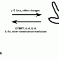
Ancillary tests differentiating benign from malignant Spitz lesion
Low AgNOR score
HMB-45 staining less intense deeply
Ki-67 staining index
Fatty acid synthase staining
Chromosome copy analysis not consistent with melanoma
Spitz nevi resemble common nevi in their architectural features, are small and symmetric, and are well circumscribed. The intraepidermal component does not usually extend beyond the dermal component, and is arranged in nests that do not become confluent or vary a great deal in size and shape among each other. Nests in Spitz nevi are vertically arranged and regularly spaced, and can have clefting artifact above the nests at the dermal-epidermal junction ( Fig. 5 ). In melanoma this retraction artifact is rare. Although junctional activity is characteristic in Spitz lesions, infiltration of cells into the upper layers of the epidermis is usually not present. There may be an occasional single cell infiltration in the upper epidermis, but pagetoid spread as seen in melanoma is not a feature. Epidermal changes include acanthosis, hyperkeratosis, and hypergranulosis. Rete ridges are usually elongated owing to the vertical orientation of the nests.
Spindle cells of a Spitz nevus are cells with fusiform shape, abundant cytoplasm, and a centrally located nucleus that has a conspicuous nucleolus. These cells are often plump. The nuclear chromatin pattern is usually finely dispersed but may be slightly vesicular. The cells are generally arranged in a fascicular pattern grouped in vertically oriented nests. The overall size of the nests and the cells within them are variable, but tend to decrease in size toward the deeper layers of the lesion.
The epithelioid cells of a Spitz nevus are large with abundant cytoplasm and have large nuclei. These cells can be round, oval, polygonal, rhomboidal, or polyangular. The nuclei can be round, oval, irregularly shaped, or multilobulated. Multinucleated forms may be present. In some cases a minority of epithelioid cells can have cytoplasmic and or nuclear contours that are very irregular. The cells can be strikingly large or have a bizarre shape. Cytoplasm sometimes has a ground-glass appearance. Melanin is typically absent or not abundant, although melanin is prominent in melanophages in the pigmented spindle variant.
Within the dermis, the cells display maturation and the nests are replaced by a single cell infiltrating pattern into the base. Kamino bodies are pale eosinophilic globules (now known to be composed of basement membrane material) that stain positive with periodic acid Schiff and trichrome, and are commonly found in the dermal-epidermal junction of Spitz nevi.
Distinction of Spitz nevus from melanoma is made by a constellation of findings taken in the context of the clinical presentation. The symmetry of the lesion, absence of significant pleomorphism, deep mitoses or pagetoid spread, and lack of extension into surrounding structures are features recognized to suggest benign biology. In contrast, melanoma is typically a large lesion that is asymmetric and poorly demarcated. Nests are variable in size, shape, and orientation, and Kamino bodies are absent. In melanoma there is lack of maturation, mitoses are prominent and present in the depth of the lesion, and there is a lack of epidermal change. Cellular type is variable in melanoma, with various degrees of pigmentation.
The diagnosis of Spitz nevus with all the typical features in the setting of young children can be straightforward. However, there is significant potential for over- or underinterpretation of pathologic findings or absence of findings because of the baseline features that can mimic melanoma. As lesions become less characteristic, determination of benign Spitz nevi from those with malignant potential based on pathology can be problematic, even for experienced practitioners.
Related posts:

Stay updated, free articles. Join our Telegram channel

Full access? Get Clinical Tree


