ANATOMY 
 Name the labeled structures in Figure 37-1.
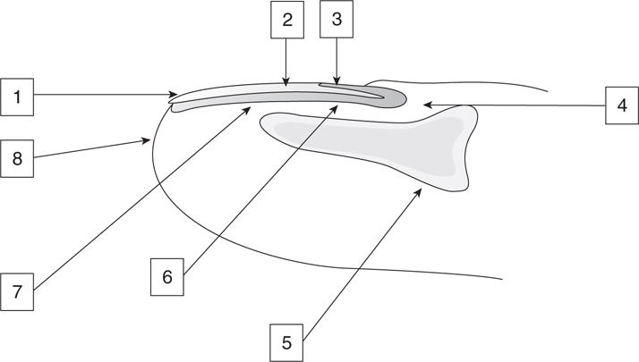
Figure 37-1 Anatomy of the perionychium.
1. Nail plate
2. Lunula
3. Eponychium
4. Nail fold
5. Distal phalanx
6. Germinal matrix
7. Sterile matrix
8. Hyponychium
 What is the perionychium?
The nail bed (composed of the sterile and germinal matrix) and surrounding soft tissues (paronychium).
 What is the hyponychium?
The junction of the nail bed (sterile matrix) and the fingertip skin, beneath the distal free margin of the nail. It consists of a keratinous plug that prevents debris from getting under the nail plate. The hyponychium also contains large numbers of leukocytes and lymphocytes and is the first barrier of defense to prevent bacteria and fungi from invading the subungual area.
 What is the eponychium?
The distal portion of the nail fold where it attaches to the surface of the nail.
 What is the paronychium?
Soft tissue around the nail and the nail folds comprises the paronychium.
 What is the nail bed?
The nail bed consists of (a) the germinal matrix on the proximal ventral floor of the nail fold and (b) the sterile matrix extending from the lunula to the hyponychium.
 What is the germinal matrix?
The germinal matrix comprises the ventral floor of the nail fold. Highly vascular and composed of germinal cells near the periosteum, the germinal matrix produces 90% of the nail volume. As the germinal cells duplicate, previously formed cells are forced toward the nail and the pressure causes the cells to flatten, elongate, and stream distally into the nail.
 What is the sterile matrix?
Part of the nail bed that extends from the lunula to the hyponychium.
 What is the nail fold? Why is it important?
The nail fold houses the proximal nail plate and is composed of the germinal matrix on the ventral floor and the portion of the nail bed that forms the cells which makes the nail shine on the dorsal roof. The patency of the nail fold is crucial for normal nail growth; hence, either the nail plate or a temporary stent should be placed to keep the nail fold open.
 What is the lunula?
The curved white opacity representing the visible, distal portion of the germinal matrix.
 What is the nerve supply to the perionychium?
Dorsal branches from the volar digital nerves.
 What is the arterial supply to the nail?
Two dorsal branches of the volar digital arteries.
 What makes up the nail plate?
Flattened sheets of anuclear keratinized epithelium densely adherent to one another.
 What changes occur in the nail plate distal to the lunula?
The cell nuclei degenerate distal to the lunula. This is the junction of the sterile and germinal matrix.
 What lies beneath the sterile matrix?
Periosteum of the distal phalanx; hence fracture of the distal phalanx is associated with a high incidence of nail bed injury.
 PHYSIOLOGY
 What produces the nail plate?
The germinal matrix produces 90% of the nail plate volume.
 What contributes to nail plate adherence?
The sterile matrix produces keratin, which thickens the nail and allows adherence of the nail plate to the bed as it migrates distally.
 What contributes to the smooth shiny surface of the nail plate?
The dorsal roof of the nail fold.
 How is the nail produced?
The nail plate is a multilayered stacked sheet of cornified cells derived from anucleate onychocytes that arise from the germinal matrix epithelium of the nail bed.
The epithelium of the germinal matrix, sterile matrix, and eponychial fold contributes to the production of the nail plate through three modes of keratinization.
The germinal matrix epithelium undergoes onychokeratinization, forming the main substance of the hardened nail plate, which is composed of stratified layers of cornified onychocytes.
The sterile matrix epithelium produces a semirigid keratin through a process known as onycholemmal keratinization. This semirigid keratin increases the overall thickness of the nail and also acts as superglue adhesive for the nail plate to maintain its adherence to the nail bed.
The eponychial fold (dorsal roof) is responsible for the external sheen of the healthy nail plate by epidermoid keratinization. The cuticle, hyponychium, and lateral nail folds also contribute, in a minor degree, to the surface epidermoid keratinization of the nail plate.
 Name five functions of the fingernail.
1. Protection of the fingertip
2. Improved pulp tactile sensation through provision of counterforce to the pulp
3. Assistance in picking up objects
4. Self-defense (scratching)
5. Regulation of peripheral circulation
 What area of the body has the highest concentration of lymphatics?
The hyponychium.
 At what rate does the nail grow?
An average of 0.1 mm/day or 100 days for complete nail growth; however, after an injury, distal growth is halted for 21 days as the proximal nail thickens.
 MEDICAL PATHOLOGY
 What is clubbing?
Exaggerated convex curvature of the nail plate.
 What conditions are thought to cause clubbing?
• Familial clubbing (idiopathic)
• Pulmonary disease—pulmonary fibrosis, sarcoidosis, cystic fibrosis
• Cardiac disease—cyanotic congenital heart disease, bacterial endocarditis
• Gastrointestinal disease—ulcerative colitis, Crohn disease, liver cirrhosis
• Cancer—thyroid, thymus, disseminated chronic myelogenous leukemia
• Other—acromegaly, pregnancy
 What is chromonychia?
Changes in nail color.
 What causes this?
Chromonychia can be induced by renal failure, subungual hemorrhage, or medications. Antineoplastic drugs frequently cause melanonychia. Drugs most commonly involved are adriamycin, cyclophosphamide, and vincristine. Chromonychia is also associated with AIDS.
 SURGICAL PATHOLOGY
 What is onycholysis?
Premature separation of the nail bed and nail plate.
Related posts:
Stay updated, free articles. Join our Telegram channel

Full access? Get Clinical Tree


