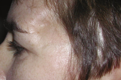
Concavity in the temporal area is often referred to as “temporal hollowing.” It reflects a deficiency in the bulk of the temporalis muscle, the temporal fat pad, and/or an underlying bone defect ( Fig. 6.1 ).
Etiology of Temporal Hollowing
When the temporal area has not been surgically violated, concavity gradually appears with senescence. It may appear in patients with low body fat, hence less temporal fat, and after massive weight loss. It may occur as a result of HIV-associated lipodystrophy and radiation-induced soft tissue atrophy. In other healthy patients, prominent adjacent skeletal contour may diminish the relative projection of the temporal soft tissues. Therefore, augmenting the contours of the temporal area can have a rejuvenating and/or a balancing effect on facial appearance.
Both aesthetic and reconstructive procedures that involve the temporal area may result in temporal hollowing. Aesthetic surgeries that access the midface via separation of the temporal fascial planes can also result in depression in the temporal fossa. Temporal depressions also occur after neurosurgical procedures during temporal or pterional craniotomy where detachment of the temporalis muscle from its origin at the temporal crest or the lateral orbital rim is required. Reattachment of temporalis muscle often yields suboptimal results, leading to significant depressions beneath the temporal line. Moreover, the deformity is exaggerated by the overlying soft tissues that are thinned out due to atrophy or posttraumatic scarring. Temporal augmentation can restore the presurgical appearance in these patients.
Treatment Options
Several procedures have been described to augment the contour of the temporal area. These include the use of various alloplastic implants, free fat grafting, injection of various absorbable and permanent filler materials, and in some instances, vascularized flaps.
Temporal augmentation with autologous fat grafting has been associated with difficult irregularities. In fact, the temporal region is one of the subunits with the lowest satisfaction rate after facial fat grafting. Patients with thick skin generally experience the best results with fat injection; however, thick skin is not a typical quality in patients with temporal hollowing due to senescence. Besides skin irregularities and edema, other severe, but rare, complications such as blindness and cerebral fat embolism have been reported. A thorough knowledge of the clinical anatomy of the temporal region is crucial for the safe performance of temporal fat grafting.
Similarly, temporal augmentation with filler materials (e.g., hyaluronic acid) produces similar contour irregularities. Adopting the technique of diluting calcium-based fillers for the dorsum of the hand, Lambros described a technique for filling the temples with highly diluted hyaluronic acid. This dilution method allows the filler to be distributed more evenly in the temporal area, resulting in a more satisfactory outcome.
Augmenting the temporal region with various alloplastic materials has been described. Contour irregularities at the interface of the implant edge and native anatomy are not uncommon. This chapter describes the senior author’s use of polymethylmethacrylate (PMMA) to fill depressions in the temporal area. In instances when no previous surgery has been performed or when the temporal area has served as a dissection plane to access adjacent areas (e.g., subperiosteal facelift), the implant material is placed beneath the temporal muscle through a limited incision in the hair-bearing scalp. When previous reconstructive surgery has been performed in the temporal area, the surgical incision scars are used to access the area of depression for placement of PMMA over the temporal muscle. The author’s techniques using PMMA to reconstruct temporal contour depressions have been reliable, durable, and relatively free of complications.
Operative Technique: Aesthetic Temporal Augmentation
Access
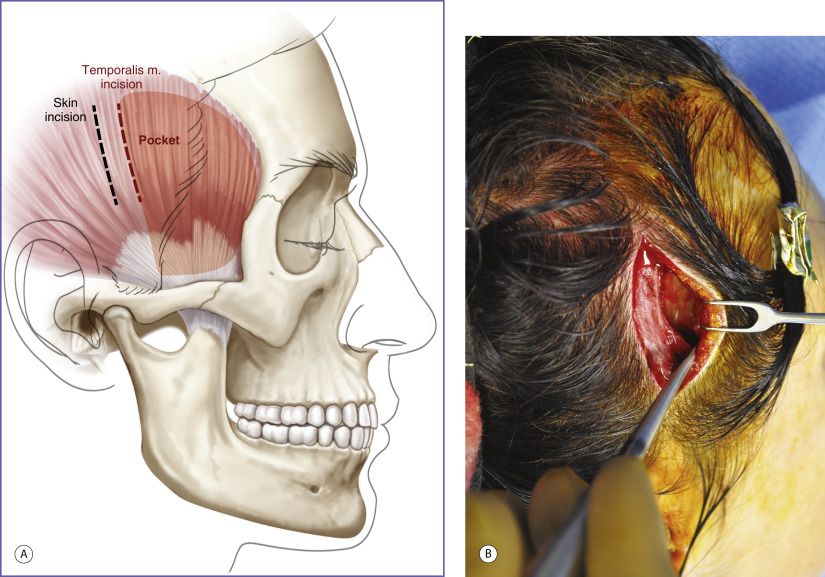
A 5- to 6-cm vertical incision is made above the helix, within the hair-bearing temporal scalp. The scalp flap is undermined anteriorly for 2 to 3 cm before the deep temporal fascia and the underlying temporalis muscle are incised along the direction of the vertically oriented muscle fibers. The temporalis muscle is split and dissection is continued to reach the temporal fossa ( Fig. 6.2A ).
Formation of submuscular pocket
After reaching the temporal bone, a space between the temporalis muscle and the underlying temporal bone is created ( Fig. 6.2B ). The dissection is done directly on the temporal bone using the coagulation mode of the electrocautery device. The dimensions of the pocket will define the extent of augmentation. Transversely, the area of undermining will extend from the lateral orbital rim anteriorly to just behind the hairline posteriorly. Vertically, the dissection extends from the temporal crest to just above the zygomatic arch. Extending the dissection beneath the zygomatic arch can result in impingement of the temporal implant on the condyle or coronoid process of the mandible.
Shaping the implant
PMMA is prepared by mixing the powdered polymer with the liquid polymer. After the mixture has achieved a putty-like consistency, it is placed in the submuscular pocket ( Fig. 6.3 ). Before the PMMA hardens, it is molded to its desired shape by manipulating the overlying soft tissues so that they project to the same level as the lateral orbital rim. Any areas of overcorrection (not uncommon laterally after molding the material to be flush with the lateral orbital rim) are revised with a contouring burr. The muscle and the temporal fascia are reapproximated and the scalp incision is closed in layers after a satisfactory temporal contour has been achieved.
Related posts:
Stay updated, free articles. Join our Telegram channel

Full access? Get Clinical Tree


