(1)
Hôpital Universitaire de Strasbourg, Strasbourg, France
Abstract
There are several semiological variants of telangiectases, some of which have a diagnostic value:
There are several semiological variants of telangiectases, some of which have a diagnostic value:
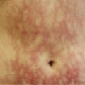
Arborising pattern (cf. Fig. 3.12a).
Stellate (cf. Fig. 3.12a).
Rectangular macules (cf. Fig. 15.27): common in sclerodermas.
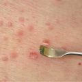
Papular (cf. Fig. 15.94): common in Rendu-Osler disease; to be distinguished from ruby spots.
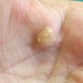
Papular and keratotic: angiokeratomas; when multiple and located in the underwear area, they command the search for Fabry disease (cf. Fig. 15.96).
Periungual or gingival, also known as “mega-capillaries” (cf. Fig. 15.21) and highly suggestive of a connective tissue disease, particularly dermatopolymyositis and scleroderma.
Associated with other lesions; present among lesions of poikiloderma or of the pearly border of basal cell carcinoma.
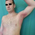
Certain telangiectases are the dermatologic manifestation of systemic diseases such as intravascular lymphoma or POEMS syndrome (polyneuropathy, organomegaly, endocrinopathy, monoclonal component, skin lesion). In patients with intravascular lymphoma, the biopsy of a telangiectasia allows to establish diagnosis, due to the fact that there is a proliferation of malignant lymphocytes that are usually easily observable within the dermal vessels. In the absence of a cutaneous lesion, the diagnosis of intravascular lymphoma which often affects the central nervous system and can mimic vasculitis and can be very difficult to establish.
Table 24.1
Main causes of telangiectases
Type of lesions
Related posts:Stay updated, free articles. Join our Telegram channel
Full access? Get Clinical Tree
 Get Clinical Tree app for offline access

|
|---|