(1)
Hôpital Universitaire de Strasbourg, Strasbourg, France
Abstract
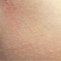
Desquamation (or scaling) is usually physiological and non-visible and corresponds to normal shedding of epidermal cell residues from the stratum corneum (nonviable cells). It sometimes becomes apparent with the formation of easily detachable scales, unlike keratosis which is firmly adherent. Scaling indicates an epidermal involvement in the pathological process. It can be present either initially or immediately after the onset of a disorder, thus being one of the primary lesions characterizing the disorder. Examples are the scaly erythematous plaques occurring in psoriasis or the scaling which always quickly accompanies erythroderma. It can also occur secondarily, such as with most drug-induced and viral exanthems. Scaly erythematous lesions are particularly common.
Desquamation (or scaling) is usually physiological and non-visible and corresponds to normal shedding of epidermal cell residues from the stratum corneum (nonviable cells). It sometimes becomes apparent with the formation of easily detachable scales, unlike keratosis which is firmly adherent. Scaling indicates an epidermal involvement in the pathological process. It can be present either initially or immediately after the onset of a disorder, thus being one of the primary lesions characterizing the disorder. Examples are the scaly erythematous plaques occurring in psoriasis or the scaling which always quickly accompanies erythroderma. It can also occur secondarily, such as with most drug-induced and viral exanthems. Scaly erythematous lesions are particularly common.
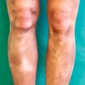
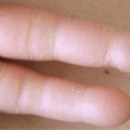
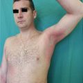
Certain types of scaling are specific and may highlight certain mechanisms and/or diseases. For example, shredding (scarlatiniform scales) indicates a brutal interruption of the stratum corneum production, which is typical of superantigen-mediated diseases. Collarette scaling is clinically characterized by a scale which is adherent in the periphery but detachable at its center, thus realizing a collarette. It is typical of pityriasis rosea (of Gibert) but also occurs in candidiasis, glucagonoma syndrome, Sneddon-Wilkinson disease, or superficial pemphigus. In psoriasis, scratching with a Brocq’s curette will initially reveal white powdered scales (“signe de la tache de bougie” in French terminology), followed by the “sign of the last removable scale” (“le signe de la dernière pellicule detachable” in French terminology), i.e., the thin suprapapillary scales being detached by the curette, and finally the Auspitz sign (“signe de la rosée sanglante” in French terminology), i.e., the formation of bleeding spots due to exposition of the hypervascularized dermal papillae. In pityriasis lichenoides, the primary lesion is an elevated pink or red papule, which rapidly turns brownish-red and is covered with dry, gray, and adherent scales. The lesion then becomes depressed, turning into a macule that retains a scale at its surface. That scale is detachable in one piece using a curette, which is typical of this disorder.
Table 41.1


Main types of scaling with some examples of associated disorders
Related posts:

Stay updated, free articles. Join our Telegram channel
Full access? Get Clinical Tree
