(1)
Hôpital Universitaire de Strasbourg, Strasbourg, France
Abstract
The distribution or extension of a dermatosis must always be emphasized. The disease may be localized (single, isolated lesion) (Fig. 10.1), segmental or regional (Fig. 10.2), diffused or generalized (Fig. 10.3), or universal (Fig. 10.4).
The distribution or extension of a dermatosis must always be emphasized. The disease may be localized (single, isolated lesion) (Fig. 10.1), segmental or regional (Fig. 10.2), diffused or generalized (Fig. 10.3), or universal (Fig. 10.4).
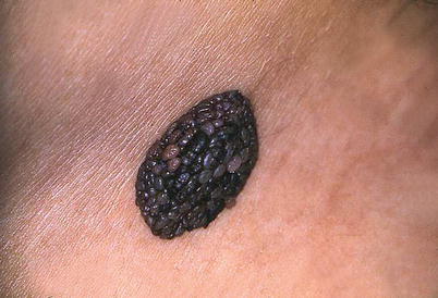
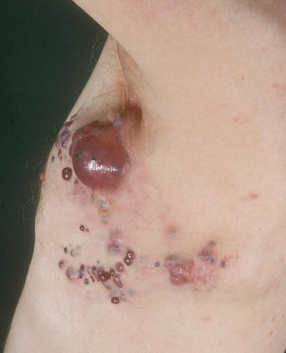
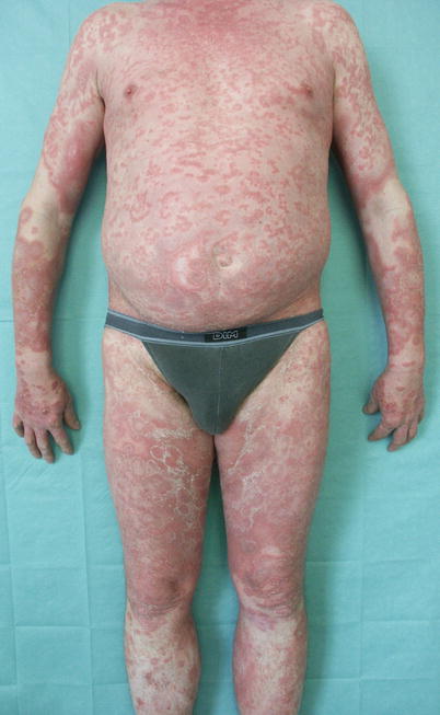
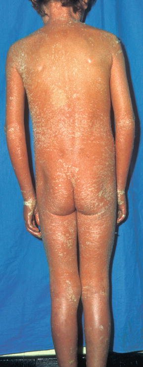
Fig. 10.1
Localized distribution. Tuberous nevus. Single lesion occurring on a very localized anatomical area. It is the case in all tumoral lesions but also in several infectious diseases. Inflammatory dermatoses such as psoriasis may be localized, segmental, diffused, or generalized
Fig. 10.2
Regional distribution. Regional cutaneous metastases in a patient with melanoma. The regional distribution may be due to several mechanisms such as the exposure of a limited cutaneous area to a pathogen, or the correspondence to an area of lymphatic drainage (as in this example), or to a metamere
Fig. 10.3
Generalized distribution. Psoriasis. In this example, every segment of the body was affected by psoriasis. However, lesions are not confluent and the distribution is not universal, as could be the case in psoriatic erythroderma
Fig. 10.4
Universal distribution. Ichthyosiform erythroderma. The entire skin is affected, including the palms and soles
Certain distributions have a remarkable aspect and their identification requires experience. A particular distribution may reflect the exposure of uncovered skin to a pathogen present in the environment, or to the sun. Therefore, these dermatoses will be located mainly on exposed areas: face, neck, neckline, and back of the hands and/or forearms (Figs. 10.5, 10.6, and 10.7). It may be difficult to distinguish a dermatitis caused by an airborne agent from a photoinduced dermatitis. Certain signs may be of help: the airborne agent will tend to accumulate in the exposed cutaneous folds such as the palpebral or retroauricular folds which are naturally photo protected. The airborne dermatitis will thus affect these areas which are not involved in photoinduced dermatitis. Likewise, the anterior cervical area located below the chin is naturally protected from sun exposure, i.e., the submental triangle, which will remain clear in photosensitive dermatitis (Fig. 10.8). Distribution also gives an indication about the underlying mechanism of a lesion: a very sharp demarcation, as if traced using a ruler, is typically found in phototoxic dermatitis (Fig. 10.9), while imprecise demarcation is characteristic of photoallergic dermatitis and photoaggravated disorders (Figs. 10.5 and 10.8).
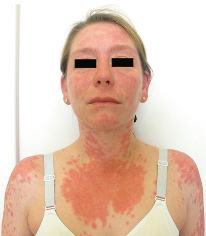
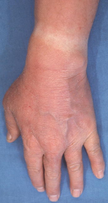
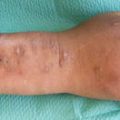
Fig. 10.5
Photodistribution. Subacute cutaneous lupus erythematosus. Lesions are predominantly located on areas exposed to ultraviolet radiation: face and neckline. Lupus erythematosus belongs to the group of photo-triggered and/or photoaggravated diseases
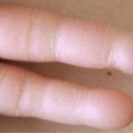
Fig. 10.6


Photodistribution. Polymorphous light eruption. Note the avoidance of the skin area protected by the watch
Related posts:

Stay updated, free articles. Join our Telegram channel
Full access? Get Clinical Tree
