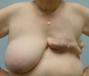
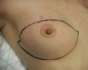
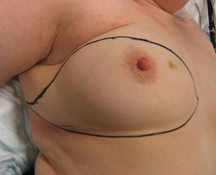
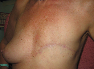
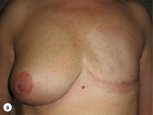
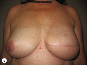
5 The patient may have risk factors for poor wound healing, which include: There are more than 4000 chemicals present in cigarette smoke, including nicotine and carbon monoxide.1 One effect of nicotine is to cause vasoconstriction of the dermal–subcutaneous vascular plexus. This has important consequences, as mastectomy flaps rely on this plexus for survival.2 As well as inducing a hypoxic state and causing vasoconstriction, smoking can lead to increased platelet aggregation, which results in the formation of tiny thromboses in capillaries. This is detrimental to wound healing, which relies heavily on blood flow in newly formed capillaries. Smokers have higher levels of fibrinogen and haemoglobin, which increase blood viscosity, which further increases the likelihood of blood clotting, and blood velocity can be reduced by up to 42% in smokers.3 The combination of decreased oxygen delivery to tissues and the thrombogenic effects of smoking, together with increased viscosity and reduced velocity, explain why wound healing in smokers is significantly impaired. The link between smoking and wound healing was first documented in the 1970s. One study of 425 patients undergoing mastectomy and breast-conserving surgery identified smoking as an independent predictor for wound infection and skin necrosis regardless of the number of cigarettes smoked.4 Another study of 716 patients having free transverse rectus abdominis myocutaneous (TRAM) flaps showed mastectomy flap necrosis, abdominal flap necrosis and abdominal hernias were significantly higher in smokers.5 This study did demonstrate a dose effect with smokers who had a history of more than a pack a day for 10 years being at increased risk of developing problems compared with smokers who had smoked for a smaller number of pack years (55.8% vs. 23.8%). One observation in this study was that delayed breast reconstruction in smokers was associated with a significantly lower rate of wound complications compared with immediate breast reconstruction in smokers. The risk of wound complications in delayed reconstructions in smokers was similar to the rate in non-smokers. Complications were also less common in women who stopped smoking 4 or more weeks before surgery. There was one small study of 108 patients investigating smoking cessation prior to surgery that was a randomised clinical trial with 40 patients in the control group and 68 patients in the interventional group.6 Patients assigned to intervention were given counselling and nicotine replacement therapy. This study showed a significant reduction in complications in the intervention group, with a reduction in both wound-related complications and the need for secondary surgery. In addition to general considerations, four questions should be answered: The all too frequently seen but completely avoidable complication of mastectomy is redundant tissue, also known as a dog ear, which is unsightly, causes difficulty with bra fitting and often chafes on the prosthesis, arm or bra (Fig. 5.1). This is a simple and very effective option to enable women with a heavy breast to wear a lighter prosthesis and feel less unbalanced (Fig. 5.2a). In some cases a woman may choose a bilateral mastectomy to achieve better overall symmetry. Figure 5.2 (a) A low mastectomy scar with contralateral reduction. (b) Delayed reconstruction with LD flap. The scar should be sympathetic to the method of delayed reconstruction planned. In most cases a low scar is best (as in Fig. 5.2a). It allows a flap-based reconstruction to be set at the inframammary fold, with the upper scar low enough to be hidden in low- neckline clothes (Fig. 5.2b). • Examine the patient sitting up to assess lateral tissue and plan the likely lateral end of scar. The predicted lateral extent of the incision can be marked. • The extent of the scar is important if radiotherapy is planned as the whole scar will be covered and can result in large volumes of tissue being treated if the scar extends a long distance posteriorly. • Mark any skin that needs to be removed over the cancer. • Decide if a second incision is required for sentinel node biopsy (usually not necessary). Most scars can be based around the inframammary fold (IMF). The incision pattern is drawn in theatre initially with a line at or just below the IMF (in women with any intertrigo the scar should be placed below this). Then with repeated upward and downward movement of the breast the planned transposition of this line on the breast skin can be marked (Fig. 5.3). In most cases the upper incision line passes a little above the areola. Attention should be paid to the degree of tension applied to the upward or downward breast movement as this represents the tension that will be exerted on the wound on closure. The upper and lower incision lines should be planned so that they meet comfortably but without excess laxity. Incisions should be planned to avoid any dog ear. To achieve this it is often best to continue the incision along the bra line laterally, curving up slightly until the upper and lower lines meet (Fig. 5.4) or, if there is doubt about how to fashion the lateral end, stop the incision at the lateral edge of the breast and fashion it once the mastectomy is complete, before closure (see comments regarding dog ear below). Transverse mastectomy scars are commonly used but rarely, if ever, can be closed without significant excess of tissue, particularly laterally. It is beholden on all surgeons to be familiar with a range of mastectomy incisions and given that there are always better alternatives, transverse mastectomy scars should be avoided. Inferior broad-based flaps can be designed to allow skin excisions in the upper pole. In breasts with a high nipple position or in cases where skin excision in the upper pole is desired, the lower incision line can be adjusted to preserve skin on the lower flap. Such modifications to the inferior skin flap should be broad based. Other scar patterns to consider in such situations include the Wise pattern or dome-shaped scar (Fig. 5.5). Lighting: A headlight is valuable and should be part of the equipment available for any breast operation. Retraction: Care should be taken with the edges of the mastectomy flaps. Sharp hooks or tissue forceps applied to dermis cause less trauma to mastectomy flaps than blunt retractors. Identifying the ‘plane’: Some would contest its existence, but there is a readily identifiable plane between the breast and subcutaneous fat that defines the dissection. That is not to say that it is obvious in every case and it may be quite irregular. The thickness of fat on mastectomy flaps varies between patients and increases further the distance from the nipple. Importantly, however, the subcutaneous vessels (extensions of the intercostal perforators) lie superficial to this plane. The plane can be defined by hydrodissection infiltrating saline (I use 1 in 1 million – others use 1 in 500 000)/adrenaline using a spinal needle, Verres needle or blunt infiltration cannula attached to a 50-mL syringe or a pressure bag (100–150 mmHg) of saline/adrenaline solution. The plane is identified as a white line after performing a skin incision before the flaps are lifted and retracted. With opposing retraction on skin and breast and light initial dissection, tissues are seen to separate at the level of the plane. Dissection then chases this white line with continued opposing retraction (with skin hook retraction on the upper flap, skin kept as straight as possible), cutting on its superficial surface. This produces a flap of uniform thickness that will be thicker in fatter women and thinner in others. Surgical tools: My preference is to use a hand-held diathermy on a fulgurate setting throughout. If using hydrodissection, scissors or a knife can be used as the plane is bloodless. Different surgeons have different preferences. Blood loss should be less than 100 mL if diathermy is used. Some prefer scissors or the knife because of concern of the ‘burn’ that results from diathermy dissection; blood loss is generally greater with scissors or the knife. Preserving the intercostal perforators: These represent the main blood supply to the mastectomy flaps once the breast tissue is removed. Their preservation is important, not just for the prevention of flap necrosis and wound problems, but also to maximise the longer-term quality of the skin. The largest tend to originate at the second or third intercostal space. These are usually encountered early in the dissection just superior to the areola and can be seen (especially in thin women) and preserved during dissection upwards and medially.
Techniques of mastectomy
tips and pitfalls
General considerations
Smoking
Considerations for simple mastectomy
Planning a mastectomy
Technique
Related posts:
 Psychosocial issues in breast cancer
 Nipple discharge and the role of ductoscopy in breast diseases
![]() The role of adjuvant systemic therapy in patients with operable breast cancer
The role of adjuvant systemic therapy in patients with operable breast cancer
 Pathology and biology of breast cancer
![]() The genetics of breast cancer, risk-reducing surgery and prevention
The genetics of breast cancer, risk-reducing surgery and prevention
![]() Oncoplastic procedures to allow breast conservation and a satisfactory cosmetic outcome
Oncoplastic procedures to allow breast conservation and a satisfactory cosmetic outcome
![]()
Stay updated, free articles. Join our Telegram channel

Full access? Get Clinical Tree


Techniques of mastectomy: tips and pitfalls