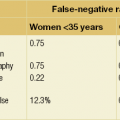
3 After breast lumps and mastalgia, spontaneous nipple discharge forms the next most common symptom, comprising 3–8% of referrals to symptomatic breast clinics.1,2 The majority of nipple discharge is benign, with up to 20% being associated with an underlying malignancy.3 Conventional methods of investigating nipple discharge include mammography, ultrasound and smear cytology, each of which have recognised limitations. Standard operations such as microdochectomy and major duct excision are undirected procedures that carry a risk of leaving undiagnosed pathology in the breast. The causes of nipple discharge are wide ranging. The majority of discharge is physiological, hormone related or results from benign breast change. A recent meta-analysis that included over 3000 women with nipple discharge demonstrated an overall incidence of underlying breast malignancy of 18.7%,3 which is higher than the figure reported in many individual series. The most common physiological nipple discharge is lactation. Ongoing milky discharge may occur up to 2 years following a pregnancy and is a normal phenomenon. Galactorrhoea can also result from prolactin-secreting pituitary adenomas, medication that influences the oestrogen, progesterone or prolactin pathways, hypothyroidism and recreational drugs such as marijuana. Commonly prescribed medical drugs that may cause nipple discharge are summarised in Table 3.1. Table 3.1 Common drugs that mimic galactorrhea and the likely underlying mechanisms of action Persistent spontaneous discharge from a single nipple orifice is usually indicative of specific pathology involving that duct. Benign intraductal papillomas account for about 80% of single-duct bloodstained nipple discharges.2,4,5 Papillomas give rise to nipple discharge of varying colour but as these structures can bleed intermittently, papillomas are often associated with blood staining. Recent bleeding may manifest as frank blood, but stagnant bloodstained fluid can be dark red, brown or black. Pathological nipple discharge is considered to be discharge arising from a single duct that is persistent (defined as more than twice per week). At age over 50 years, the presence of blood in the discharge and the presence of a clinical lump increase significantly the risk of associated malignancy,3,6 and it is recommended that these patients are fully investigated by conventional imaging techniques. Normal imaging should direct further investigation as appropriate and consideration of diagnostic surgical excision. However, a recent meta-analysis found a higher incidence of underlying malignancy in patients with bloodstained nipple discharge compared to those with non-bloody discharge (of any colour description; odds ratio (OR) = 2.27, 95% confidence interval (CI) = 1.32–3.89) or when compared to those with serous discharge alone (OR = 2.49, 95% CI = 1.25–4.93).3 It is therefore recommended that all patients with bloodstained nipple discharge are investigated fully. Mammography is indicated in women over the age of 35 years and may demonstrate a mass or microcalcification in association with nipple discharge of malignant aetiology. Even in younger women with bloodstained discharge, mammography can demonstrate calcification in women with widespread ductal carcinoma in situ (DCIS). Other mammographic features may also be evident in patients with benign nipple discharge. Duct ectasia may occasionally be visible on mammography. The sensitivity of mammography in detecting pathology in nipple discharge is 57.1% (positive predictive value (PPV) of 16.7% and negative predictive value (NPV) of 91.4%).7 In a study of 306 patients with nipple discharge who had normal mammography and ultrasound, 10% were subsequently found to have underlying malignancy.7 Ductography is less widely used but is an investigation that gives clear delineation of the breast anatomy. A small amount of radio-opaque contrast is instilled into a cannulated nipple duct. Accurate detection of small filling defect(s) caused by papillomas and duct narrowing or obstruction from malignant change can be recognised. However, ductography does not provide a tissue diagnosis and the precise position of the area of interest within the breast is not usually available to the surgeon, although it is possible to target the area of abnormality with a wire localisation procedure. The use of MRI to evaluate the ductal tree is gaining interest but is not a standard investigation of nipple discharge. In a comparative study of 163 patients with nipple discharge who had normal mammography and ultrasound, ductography was found to have a PPV of 19% and an NPV of 63%. MRI was performed in 52 patients and found to have a PPV of 56% and an NPV of 87%.7
Nipple discharge and the role of ductoscopy in breast diseases
Introduction
Causes of nipple discharge
Mechanism of action
Medication
Dopamine receptor blockade
Antidepressants:
Selective serotonin reuptake inhibitors (citalopram, fluoxetine, paroxetine, sertraline)
Tricyclic antidepressants
Antipsychotics:
Risperidone
Butyrophenones (haloperidol)
Phenothiazines (chlorpromazine)
Thioxanthenes (chlorprothixene, flupenthixol)
Anti-emetics:
Metoclopromide
Domperidone
Dopamine depletion
Methyldopa
Reserpine
Monoamine oxidase inhibitors
Inhibition of dopamine release
Codeine
Heroin
Morphine
Histamine receptor blockade
Cimetidine
Famotidine
Ranitidine
Stimulation of breast tissue and lactotrophs
Oral contraceptives
Mechanism unknown
Verapamil
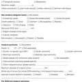
Assessment


Related posts:
 Psychosocial issues in breast cancer
 Litigation in breast surgery
![]() The role of adjuvant systemic therapy in patients with operable breast cancer
The role of adjuvant systemic therapy in patients with operable breast cancer
 Pathology and biology of breast cancer
![]() The genetics of breast cancer, risk-reducing surgery and prevention
The genetics of breast cancer, risk-reducing surgery and prevention
![]() Oncoplastic procedures to allow breast conservation and a satisfactory cosmetic outcome
Oncoplastic procedures to allow breast conservation and a satisfactory cosmetic outcome
![]()
Stay updated, free articles. Join our Telegram channel

Full access? Get Clinical Tree


Plastic Surgery Key
Fastest Plastic Surgery & Dermatology Insight Engine
