and Emir Q. Haxhija2
(1)
Department of Plastic Surgery and Burns, Institute for Mother and Child Health Care of Republic Serbia, University of Belgrade, New Belgrade, Serbia
(2)
Department of Pediatric and Adolescent Surgery, Medical University Graz, Graz, Austria
Keywords
SyndactylyChildrenTreatment9.1 Introduction
Syndactyly is defined as an abnormal interconnection between adjacent digits as a failure of differentiation of the mesenchymal structures [1, 2]. In normal anatomy, the distal end of the web lies on the palmar side roughly at the midlevel of the proximal phalanx [3]. Syndactyly can appear isolated, associated with other deformities in the upper or lower extremity, with polydactyly and/or clefting, or as part of a syndrome (Poland’s, Apert’s syndrome) [3–9]. Numerous techniques have been described for correction of syndactyly [7, 9].
9.2 Embryology
Around the day 26 (4 weeks after fertilization) the upper limb bud appears as an oblong ventrolateral bulge on the body wall between somites 9 and 12 [4, 5, 7, 10]. The emerging limb bud is composed of somatic lateral plate mesoderm covered by ectoderm [1, 4]. Subsequent limb bud growth and differentiation are controlled by distinctive regions—signaling centers: AER, apical ectodermal ridge (proximodistal growth); Wnt, Wingless type (dorsoventral growth); and ZPA, zone of polarizing activity (anteroposterior-radioulnar growth) [1, 3, 4, 7]. Digits are recognizable at 41–43 days and fully separate at 53 days [3, 10]. The process of apoptosis is needed for the separation of the fingers (it’s mediated by bone morphogenetic protein 4—BMP-4) [3, 5]. These anomalies probably occur because of differentiation disturbances in the developing hand plate [3, 7].
9.3 Epidemiology
Syndactyly is one of the most common congenital hand malformations with an incidence of 1–2 per 2000 live births (most common in Caucasians) [2–4, 7–9]. About 50% of patients with syndactylia have bilateral involvement, males are more affected than females, and it is familial in 15–40% of cases [3, 4, 6, 7]. In isolated syndactyly, the third and fourth web are the most commonly involved [3, 6, 8].
9.4 Classification
Syndactyly can be classified as simple involving soft tissue only (incomplete that does not include fingertips and complete that does include fingertips), complex (with distal bone union), and complicated (with more than only distal bone fusion) (Figs. 9.1 and 9.2) [1, 5, 6, 8]. The formal syndactyly classification of Temtamy and McKusick with five distinct subtypes has been expanded up to nine types and numerous subtypes [7].
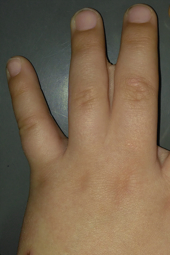
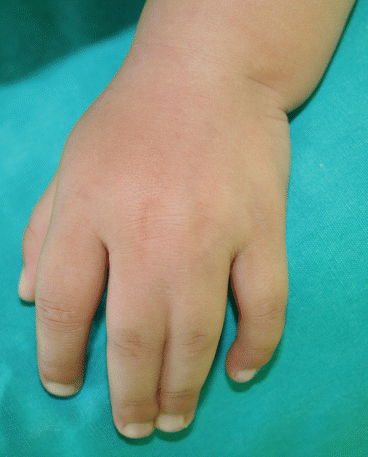
Fig. 9.1
Incomplete simple syndactyly of the third interdigital space
Fig. 9.2
Complete simple syndactyly of the third interdigital space
Incomplete syndactyly is presented as the fusion of the fingers proximal to the distal phalanx [1, 3]. In the complete form of syndactyly, the nails can be separated with full pulps of the affected fingers, or there are conjoined nails, and when fingers are of unequal length, the longest finger will tend to bend more during growth [1, 3, 5–8].
Complex syndactyly (with distal bone fusion) can involve only two fingers when it is recognized by a tapered distal end with inward rotation of the fingers and abnormally ridged or confluent nails or more fingers can be involved when they are flat to very cupped with anomalous nails, abnormal bones, and joints (Fig. 9.3a, b) [3, 6]. Complicated syndactyly is a broad category characterized by abnormal osseous abnormalities including fusions, rudimentary bones, missing bones, abnormal joints, and sometimes crossbones [1, 5, 6]. Thumb-index syndactyly treatment is more complicated than finger syndactyly, and it is treated differently from other fingers [7].

Fig. 9.3
Complex syndactyly of the hand in Apert’s syndrome: (a) clinical appearance; (b) radiography of both hands revealing fusion of distal bones
9.5 Patient Presentation
Syndactyly can be present in a large variety of forms, and that is why good preoperative planning and thorough discussion with family are important [1–8]. The fingers can be normal or anomalous, and the number of affected fingers can differ, as well as the nature of involvement [1–7, 11–17]. In complicated syndactylies, careful assessment of each individual finger is necessary before the surgery [1, 3, 16]. Radiography of the affected hand should be performed to exclude skeletal deformities. Timing of surgery depends on the fingers involved and whether the syndactyly is cutaneous or not [1–8].
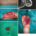
Early indications for surgery are thumb-index syndactyly, syndactylies between fingers of unequal length, and complex or complicated acrosyndactyly in order to prevent bone and joint deformities and asymmetric growth (Figs. 9.4a–c and 9.5a, b) [3, 6, 7].
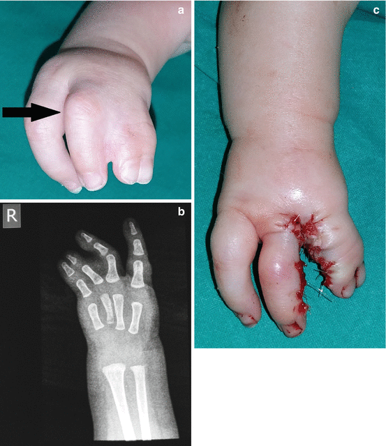
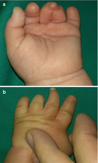
Fig. 9.4
Early indications for syndactyly correction: (a) flexion deformity of the middle finger caused by the fourth finger; (b) radiographic finding; (c) surgical treatment
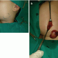
Fig. 9.5
Fenestrated syndactyly: (a) syndactyly of the distal part of the fingers with preoperative view; (b) postoperative result
9.6 Treatment/Surgical Technique
The release of syndactyly by classical technique implicates separation of the conjoined skin and subcutaneous tissue preserving integrity of the neurovascular bundles [4, 9, 12]. Techniques for separation of fingers include commissure reconstruction, digital incisions, and ways to outcome the lack of skin (Fig. 9.6a–h) [4, 6, 8]. The nail fusion can be corrected by opposing Z-flaps as described by Buck-Gramcko [4]. For safety reasons, two adjacent complete syndactylous fingers are not separated at the same time as vascular anatomy can be different [3, 4, 6]. In bilateral involvement, both hands should be operated at the same time [6].
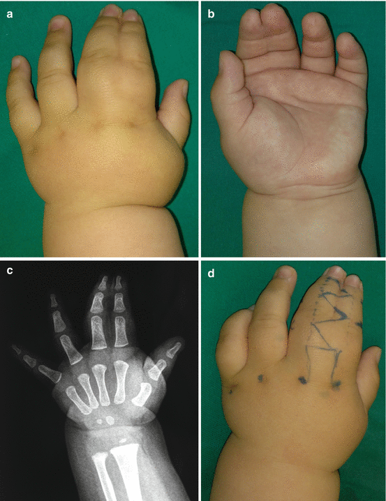
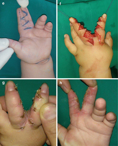
Fig. 9.6
Surgical treatment of syndactyly: (a) preoperative dorsal view; (b) preoperative palmar view; (c) preoperative radiography; (d–f) incision markings and incisions; (g, h) result
9.6.1 Digital Incisions
Separation is carried out via dorsal and standard volar zigzag incisions, creating interdigitating flaps distal to the flap for web reconstruction and providing coverage at the proximal IPJ [4, 14]. The triangular flaps can either be fully or partially interdigitated depending on the extent of the skin shortage [12, 15]. Some authors used modified zigzag incisions as longer, narrower, angled flaps or rectangular flaps joined with straight-line incisions [8, 14–16]. Digital defatting is also very important in surgical procedure to decrease the digital volume [8, 14]. It should be performed carefully, under loupe magnification, and there will be no consequences in flap vascularization or digital contour [14].
Related posts:
Stay updated, free articles. Join our Telegram channel

Full access? Get Clinical Tree


