Surgical Treatment of Rectocele and Perineal Defects

20 Surgical Treatment of Rectocele and Perineal Defects


Pelvic organ prolapse is very common. Annually, approximately 200,000 women undergo prolapse surgery in the United States. Approximately three fourths of women with prolapse have a rectocele. Although rectocele repair has been commonly performed for over a century, the long-term functional and anatomic outcomes and ideal procedure have not been determined. This chapter will review the anatomy, pathophysiology, evaluation techniques, and surgical management of rectoceles and perineal body defects.


ANATOMY AND PATHOPHYSIOLOGY


The terminology of the anatomic tissue that is present under the vaginal epithelium has been the subject of debate for most of the past century. The term fascia was introduced by Emmet in 1883. The histology of the apical portion of the posterior vaginal wall consists of mucosa (which includes the epithelium of the posterior wall and the lamina propria), a superficial and deep muscularis layer, and adventitia. This fibromuscularis has been named rectovaginal fascia and perirectal fascia, perhaps giving the surgeon an illusion of sturdier tissue than is actually present. Comparisons of the histology of women with and without prolapse have shown that the smooth muscle content of the posterior vaginal wall of women with prolapse is disorganized and significantly reduced in comparison to women without prolapse.


Prolapse of the posterior vaginal wall may be secondary to the presence of an enterocele, sigmoidocele, or rectocele, or a combination of these entities. A rectocele is an anterior protrusion of the rectal wall to the posterior vaginal wall. The rectovaginal space exists between the vaginal tube and the rectum. This potential space, occupied by areolar tissue, allows the vagina and rectum to function independently of each other. Support of the posterior vaginal wall is provided by a complex interaction of the integrity of the vaginal tube, the connective tissue support, and muscular support of the pelvic floor. DeLancey (1996) divided the connective tissue support of the vagina into three levels. All three levels of support should be evaluated and addressed during surgical management of the posterior vaginal wall.


At level I, the apical portion of the posterior vaginal wall is suspended and supported primarily by the cardinal-uterosacral ligaments. This mesentery of support originates at the sacrum and the pelvic sidewalls and inserts onto the posterior cervix and upper vagina. With normal support, the apical posterior wall of the vagina is dorsally directed to lie upon the rectum in a horizontal fashion overlying the levator ani muscles. With increases in abdominal pressure, the vaginal tube is closed top-to-bottom and supported by the pelvic floor muscles.


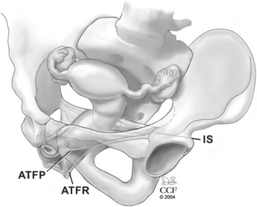
Level II includes the support for the middle half of the vagina. This support is provided by the endopelvic fascia attaching the lateral posterior vaginal wall to the aponeurosis of the levator ani on the pelvic sidewall. Most of the fibers of the endopelvic fascia connect the lateral edge of the vaginal tube to the pelvic sidewall. Very few of the fibers actually run like a sheet from sidewall to sidewall. This lateral attachment, the arcus tendineus fasciae rectovaginalis, is dorsal to the arcus tendineus fasciae pelvis for the distal half of the posterior wall (Fig. 20-1). The sidewall attachment of the posterior vaginal wall converges with the sidewall attachment of the anterior wall approximately midway in the vaginal canal. The proximal half of the posterior vagina is supported by bilateral endopelvic attachments to the arcus tendineus fasciae pelvis.


The role of the perineal body is to resist caudally directed forces by the rectum and to provide a physical barrier between the vagina and rectum. The perineal body is thicker (approximately 3 cm long) and more defined in women. It includes interlacing muscle fibers of the bulbospongiosus, transverse perinei, and external anal sphincter. The perineal body is anteriorly attached to the vaginal epithelium and muscularis of the posterior vaginal wall. Laterally, the perineal body is attached to the ischiopubic rami through the transverse perinei muscles and the perineal membrane. Anteriorly, the perineal membrane spans the anterior half of the pelvic outlet and is comprised of dense fibromuscularis. The perineal body extends cranially in the posterior wall of the vagina to approximately 2 to 3 cm proximal to the hymenal ring. This dense, fused level of support represents level III. Posteriorly, the perineal body includes the anterior portion of the external anal sphincter and its attachment to the longitudinal fibrous sheath of the internal anal sphincter. The perineal body is suspended by and attached to the puborectalis muscle. Interruption in the support of the perineal body will allow the posterior vaginal wall, perineal body, and the distal portion of the anterior rectal wall to descend with increased abdominal and rectal pressure.


The puborectalis also provides a sling of support for the vaginal tube. This sling leads to an angulation of the midposterior wall of approximately 45 degrees from vertical. The proximal portion of the vagina lies upon (and is supported by) the pubococcygeus and iliococcygeus muscles. The puborectalis helps close the potential space of the vagina and close the levator hiatus. With a healthy pelvic floor, little stress and strain are placed on the connective tissue support system.


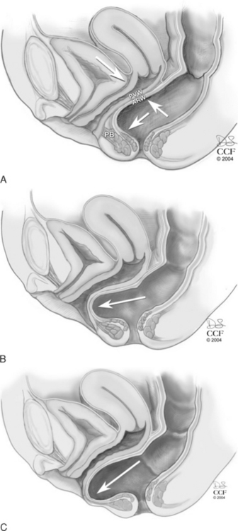
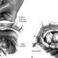
The levator hiatus has been shown to be larger in women with prolapse than in those with normal support. In a woman with an intact pelvic floor, the puborectalis is in a chronic state of contraction. This contraction closes the vaginal canal, and the anterior and posterior vaginal walls are in direct apposition. With defecation, the increased pressure placed on the posterior vaginal wall is equilibrated by the opposing anterior vaginal wall, and minimal stress is placed on the endopelvic fascial attachments (Fig. 20-2, A). If there is muscular and/or neurologic damage to the puborectalis, the levator hiatus widens and the vaginal canal opens. The increased rectal pressure and distension associated with defecation places strain on the endopelvic fascial attachments and the fibromuscularis of the posterior vaginal wall and can result in rectocele and perineal descent (Fig. 20-2, B,C).


Any condition or event that damages the support of the posterior vaginal wall can lead to prolapse. Vaginal delivery, particularly in the occipitoposterior position, is associated with an increased risk for posterior vaginal wall and perineal body trauma. Magnetic resonance images in the postpartum period show changes in intensity within the levator ani muscle. These changes likely reflect the recovery process following neurologic or muscular damage related to childbirth. Aging may also affect the levator ani muscles, leading to muscle atrophy and devascularization.


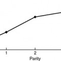
Chronic strain and constipation have been associated with (but do not necessarily cause) rectocele, perineal descent, and fecal incontinence. With chronic straining, a stretch is placed in the pudendal nerve and the nerve to the levator ani muscle. Meschia et al. (2002) found that fecal incontinence was more prevalent in women with a rectocele that extended beyond the hymen (31%) than in women with prolapse inside the hymenal ring (19%). Increasing body mass index has been strongly associated with incident rectocele but not with prolapse of other areas of the vagina (cystocele or uterine prolapse).


Pelvic surgery can predispose a woman to develop prolapse. Alterations of the connective tissue support, and injury to the innervation and vascularization to the pelvic floor muscles, occur with pelvic surgery. Alterations of the axis of the vagina may increase the forces placed on the connective tissue supports. Overelevation of the anterior vaginal wall, as with a retropubic urethropexy or needle suspension procedure, alters the distribution of force on the vaginal walls and can open the posterior wall to the development of an enterocele or rectocele.


EVALUATION


History


Many women with a rectocele or a perineal body defect are asymptomatic. However, women may complain of symptoms associated with prolapse, such as bulging of the vagina and pressure, which worsens by the end of the day and improves when lying down. Sexual dysfunction also occurs in some women with prolapse. A woman may reduce sexual activity due to the discomfort of the prolapse or the embarrassment of the urinary or anal incontinence, which may accompany her prolapse. A woman with a perineal body defect, which leads to a widened genital hiatus, may describe loss of sensation for herself and her partner during intercourse.


Defecatory dysfunction and pelvic organ prolapse are both common in women. It is often difficult to determine whether there is a causal link between the posterior wall prolapse and defecatory dysfunction or whether they are separate, concurrent disorders. Many women state that they are constipated. It is important to determine what a woman means when she states that she is constipated. Constipation includes excessive straining; hard, lumpy stools; splinting; feeling of incomplete emptying; and infrequent stools. Infrequent defecation is not likely related to a rectocele and may require additional evaluation. Women with a large rectocele may trap stool within this rectal pocket, leading to feeling of incomplete emptying, which can result in soiling. Splinting, or placing manual pressure in the vagina, over the rectum, or on the perineum to reduce the prolapse and facilitate emptying of the rectum, is commonly described.


Anal incontinence is commonly seen in patients with posterior wall and perineal body defects. Many women are reluctant to initiate the conversation about anal incontinence due to embarrassment, so it is important to ask about accidental loss of solid or liquid stool or gas.


An important part of the history to obtain from your patient is an understanding of her management desires. If the patient requires and is willing to undergo surgical management, having her express her expectations of surgery can be illuminating.


Physical Examination


The patient is generally examined in the dorsal lithotomy or semirecumbent position. An excellent correlation exists in the evaluation of prolapse between the supine and standing positions in women performing maximal Valsalva maneuver. If the prolapse observed in the lithotomy position does not recreate the degree of prolapse that the patient described, a standing examination should be performed. However, it is physically more difficult to make measurements of the prolapse in this position.


To stage the severity of prolapse the posterior vaginal wall is visualized with the posterior blade of a bivalve speculum or a Sims speculum. The retractor elevates the anterior wall and reduces any uterine or apical prolapse. The patient is asked to increase abdominal pressure with a Valsalva maneuver or cough. The Pelvic Organ Prolapse Quantification (POPQ) system is a standardized, validated tool for measuring and staging pelvic organ prolapse (see Chapter 5). Measurements of the posterior vaginal wall are documented at maximal strain, 3 cm proximal to the hymen (Ap), at the most dependent portion of the posterior vaginal wall proximal to this mark (Bp), and at the vaginal cuff (C) or cul-de-sac, if the uterus is present (D). The genital hiatus (GH) and perineal body (PB) are measured with the patient straining. Evaluation for and staging of concurrent anterior wall and apical prolapse should be performed.


The perineal body should be evaluated for descent. It may be difficult to measure perineal descent, but documentation of its presence or absence can be helpful in planning your surgery. Descent of the perineal body occurs with a lack of continuity from the suspensory support at the apex (level I) to the perineal body (level III). It may also occur because of a mass effect of the rectum or small bowel herniating into the perineal body, a perineocele. Perineal descent has also been associated with fecal incontinence. Nerve stretch and subsequent neuromuscular damage is one of the proposed mechanisms of fecal incontinence.


Perform a careful rectovaginal examination evaluating the posterior vaginal wall and perineal body. If the area of anterior rectal wall bulging and the posterior wall prolapse is the same, this provides clinical confirmation of the presence of a rectocele. Palpation of loops of small bowel confirms an enterocele; a sigmoidocele is diagnosed if sigmoid colon is palpated. Performing a rectovaginal examination in the standing position may increase the detection of an enterocele by allowing the bowel to enter the enterocele sac. This search for an enterocele should continue in the operating room. If an enterocele is missed, posterior wall prolapse may persist following surgical management. Elevating the rectal finger up to the posterior vaginal wall helps identify an area with less support, although the clinical examination has not been shown to be accurate in comparison to the surgical identification of site-specific defects of the posterior vaginal wall. Last, pressure on the posterior wall of the vagina, directed downward toward the rectum, may facilitate identification of rectal prolapse or intussusception.


Because anal incontinence commonly occurs with rectocele, assessment of the anal sphincter should be performed. This includes evaluation of anal tone, squeeze, and symmetry. If a symptomatic woman is found (or suspected) to have a disrupted anal sphincter on examination, further testing is indicated.


A focused neurologic examination includes evaluation of sensation, motor function, and reflexes of sacral nerves 2–4. The patient is asked to discriminate between sharp and dull on the perineum. Assess pelvic floor muscle strength with the patient contracting and relaxing the pelvic floor muscles around the examiner’s fingers. Reflex testing includes the bulbocavernosus reflex and anal wink (see Chapters 6 and 11.


Diagnostic Tests


A number of imaging techniques have been used to evaluate rectoceles. Defecography provides a two-dimensional view of the efficiency of rectal emptying and quantification of rectal parameters. The size of the rectocele is determined by measuring the distance between the line of the anterior border of the anal canal and the maximal point of the bulge of the anterior rectal wall into the posterior vaginal wall. Rectoceles have been described in nulliparous women. Anything less than 2 cm is considered normal, whereas a rectocele is considered large if the anterior rectal wall protrudes more than 3.5 cm. Contrast in the small and large bowels may also reveal the presence of an enterocele, sigmoidocele, or perineocele. Rectal intussusception and perineal descent may also be identified, although the clinical significance of rectal intussusception has not been determined. The dynamic nature of the study allows for insight into the defecation process. Retention of more than 10% of the barium following defecation is referred to as barium trapping. This examination is done in an artificial environment, which may make the patient more prone to incomplete emptying.


A situation in which defecography may convert a surgical procedure to one managed conservatively is the identification of pelvic floor dyssynergia. Defecation is effective through the coordination of relaxation of the levator ani and external anal sphincter and contraction of the colon. If the puborectalis or external anal sphincter is paradoxically contracted during defecation, this situation might respond to more conservative measures, such as the use of enemas or biofeedback. An electrophysiologic analysis of the puborectalis may also lead to this diagnosis. Another situation in which defecography might influence treatment is the identification of a sigmoidocele; the surgical approach to prolapse management might also include a sigmoid resection.


Rectoceles that retrain contrast tend to be larger than those that do not. However, fluoroscopic evidence of barium trapping does not relate to patient symptoms. In the symptomatic, elderly population, Savoye-Collet et al. (2003) found no association between the abnormalities demonstrated by defecography and symptoms. Defecography performed following surgical management of rectoceles has generally shown a reduction in the size of the rectocele and improvement in emptying. The limitations of defecography include that it requires special equipment, exposes the patient to radiation, does not show the rectum and adjacent soft tissue structures simultaneously, and is uncomfortable and poorly accepted by patients.


Dynamic magnetic resonance imaging (MRI) provides high-quality images of the pelvic soft tissues and viscera. MRI is noninvasive and does not require ionizing radiation or significant patient preparation. However, poor correlation exists between MRI grading of prolapse and clinical staging. To accomplish a dynamic MRI at most facilities, the woman is placed in the dorsal supine position with her legs together. MRI defecography has also been performed in the dorsal supine position with a sonographic transmission gel placed in the rectum and vagina. Images are obtained resting and while performing a Valsalva maneuver and with evacuation. However, during a Valsalva maneuver in this position, the true extent of the prolapse may not be exhibited because this is not a normal position for defecation and may not simulate that woman’s ability to defecate. An upright evaluation has been described but requires an open configuration MRI unit that is available in only a few medical centers. The limitations of this method of imaging include a lack of standardization of grading of prolapse, high cost, and relatively limited availability.


At this time, there is a lack of a standardized method of establishing a radiologic diagnosis of rectocele. Clinical examination has good sensitivity for the detection of a rectocele; therefore, radiologic confirmation of the presence or absence of a rectocele is not worthwhile. Although defecatory dysfunction is common in women with prolapse, the extent of the prolapse does not necessarily correlate with the extent of bowel symptoms. If the woman’s primary complaint is defecatory dysfunction or fecal incontinence and not a bulge, surgical correction of the rectocele or perineal body defect may not correct her symptoms. Ancillary testing is then pursued based on the woman’s complaints. Validated functional and quality-of-life questionnaires are now available. These may be performed preoperatively and postoperatively to provide a standardized method of evaluating the surgical outcomes. The patient’s preoperative symptoms and surgical goals will guide the provider in the selection of additional testing.


A woman who describes lifelong infrequent bowel movements (less than one per week) and an absence of a daily urge to defecate is unlikely to be cured of her constipation with a rectocele repair. A colon transit study may be helpful in identifying patients with slow-transit constipation. Dietary modifications, including fiber and laxatives, should be encouraged in any woman whose main complaint is constipation (see Chapter 25).


In women with symptoms of anal incontinence, an evaluation and attempt at medical management should be performed before rectocele repair. An endoanal ultrasound provides anatomic detail of the integrity of the external and internal anal sphincters; EMG study of the external anal sphincter can provide neurologic information.

Stay updated, free articles. Join our Telegram channel

Mar 11, 2016 | Posted by in Reconstructive surgery | Comments Off on Surgical Treatment of Rectocele and Perineal Defects

Full access? Get Clinical Tree

Get Clinical Tree app for offline access