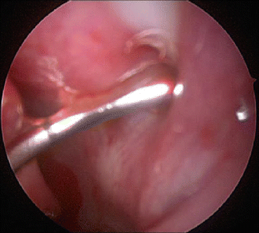
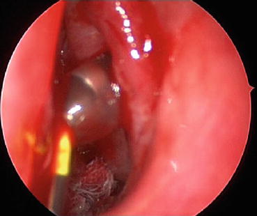
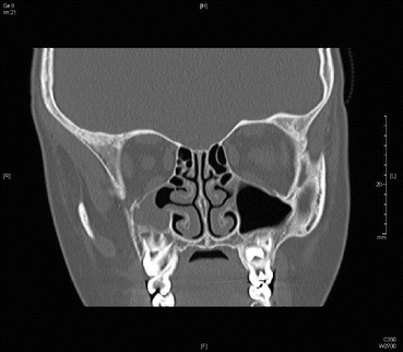
Fig. 30.1
The sinus balloon is passed over the guidewire
Use markers on balloon to ensure proper placement (Fig. 30.2).


Fig. 30.2
Markers are used on the sinus balloon to ensure proper placement
Inflate the balloon to 12 atmospheric pressure with special inflation device for the Acclarent balloon, or just use the special inflation device in the Entellus package.
Irrigation/wash of sinus as needed.
Remove balloon and wire and then remove guide.
Confirm dilatation of ostium with direct visualization.
MeroGel packing (Medtronic, Jacksonville, Florida) is typically sufficient.
Pitfalls and Tips
Create an accessory ostium.
Strip the mucosa posteriorly, thus collapsing the whole lining anteriorly.
Significant injury to ostium and uncinate process to the extent that an antrostomy may be needed.
Guide catheter/probe is introduced tip up (personal preference for children).
Hold tension on balloon while inflated to prevent inflated balloon from slipping inside/outside the sinus (Fig. 30.3).

Fig. 30.3
Hold tension on the sinus balloon while inflated to prevent the inflated balloon from slipping inside/outside the sinus
Postoperative Care
Child is discharged after a period of observation.
Oral antibiotics are given for 7 days postoperatively.
Avoid nose blowing.
Follow up in 2 weeks.
Contraindications to Balloon Catheter Dilatation in Children
Previous sinonasal surgery in target ostia
Cystic fibrosis
Extensive sinonasal osteoneogenesis
Sinonasal tumors or obstructive lesions
History of facial trauma that distorts sinus anatomy and precludes access to the sinus ostium
Ciliary dysfunction
It is also helpful to have experience with balloons in adults prior to working on children.
Endoscopic Sinus Surgery
The procedure is indicated for complicated rhinosinusitis, nasal polyposis, fungal sinusitis, and children with CRS after failure of medical treatment. A CT scan prior to surgery is mandatory. The CT scan will allow for evaluation of anatomy and extent of disease. CT scan should be available in the operating room at all times during the procedure. Image-guided surgery can be performed for cases with polyps, complicated sinusitis, or revision cases. A special consideration should also be given for cystic fibrosis patients. The following are required instrumentation for the procedure:
1.
0°, 30°, and 70° rigid scopes preferably 4 mm in size
2.
Straight and upturned Blakesley forceps of different sizes
3.
Straight and upturned through-cutting forceps
4.
Double-blind ostium seeker
5.
Short and long curved antrum cannula
6.
Right-side and left-side backward punch cutting forceps
7.
3, 5, and 7 French Frazier suction tubes
8.
Cottle elevator
9.
If needed, powered microdebrider with aggressive 2.9 and 4 mm blades
10.
Additional instruments as deemed appropriate by the surgeon for frontal sinus and sphenoid surgery
Prior to start of the procedure, the following preparations are necessary:
1.
ESS in children is done under general anesthesia.
2.
Oxymetazoline 0.5 % solution-impregnated pledgets are used for topical vasoconstriction.
3.
Injection of the middle turbinates, the uncinate processes, bulla ethmoidalis, and septum adjacent to the middle turbinates with 1 % lidocaine solution with 1:100,000 epinephrine.
4.
Child is placed supine with the head of the table slightly elevated.
5.
The surgeon should be facing the patient with the monitor facing the surgeon.
We routinely give the patient Decadron 0.15 mg/kg IV bolus.
Procedure
1.
The 0° 4 mm scope is introduced into the nasal cavity after the pledgets have been removed.
2.
If more injection is needed, it can be performed at this stage.
3.
Using the Cottle elevator the middle turbinate is mediatized until the uncinate process and bulla are visualized.
4.
Using the seeker the area of the maxillary sinus ostium is found and the ostium palpated. This can be performed in a retrograde or anterograde manner. I prefer the anterograde technique because it prevents the posterior maxillary mucosa from stripping.
5.
The ostium can then be widened posteriorly by removing the inferior edge of the uncinate process with a straight cutting forceps. For retrograde technique the right-sided backbiter can be used to remove the uncinate process anteriorly. Care should be taken not to injure the nasolacrimal duct with this technique.


