© Springer International Publishing Switzerland 2015
Pete S. Batra and Joseph K. Han (eds.)Practical Medical and Surgical Management of Chronic Rhinosinusitis10.1007/978-3-319-16724-4_1717. Oral and Topical Antifungals
(1)
Department of Otorhinolaryngology, Academic Medical Centre, Meibergdreef 9, Amsterdam, NH 1100DD, The Netherlands
Keywords
Oral AntifungalsTopical antifungalsCRSAmphotericin BAllergic fungal rhinosinusitisImmunotherapyKey Take-Home Points
The role of fungi in various forms of CRS remains to be defined.
The immunocompetence of the patient is of great importance, as invasive fungal rhinosinusitis is usually found in immunosuppressed patients.
Antifungal drugs and immunotherapy may have a role as adjuvant therapy in allergic fungal rhinosinusitis, but evidence is poor to support recommendations.
There is no indication for antifungal treatment in CRS with or without nasal polyps.
Introduction
Allergic fungal rhinosinusitis (AFRS) as a distinct clinical entity was first reported in 1976 [1]. In the 1990s it was suggested that fungus could be an important contributor to chronic rhinosinusitis [2]. With new culture techniques, it was possible to culture fungus like Alternaria and Aspergillus in most patients with CRS. It was hypothesized that fungus might play a causal role in the disease.
For causality, however, we need Koch’s postulates. These four criteria are designed to establish a causative relationship between a microbe and a disease:
1.
The microorganism must be found in abundance in all organisms suffering from the disease but should not be found in healthy organisms.
2.
The microorganism must be isolated from a diseased organism and grown in pure culture.
3.
The cultured microorganism should cause disease when introduced into a healthy organism.
4.
The microorganism must be reisolated from the inoculated, diseased experimental host and identified as being identical to the original specific causative agent.
The second of Koch’s postulates was shown. However, quite soon after the finding of fungus in patients with CRS, the first reports evolved showing that fungus was also found in healthy individuals therewith defying the first Koch’s postulate. Also the third postulate about introducing fungus in a healthy individual should create CRS could not be fulfilled and finally also the last postulate could not be fulfilled. This proved that fungus as such cannot cause CRS [3–5].
However, much more interesting than a pure causal relationship might be the option that fungus is a disease modifier within CRS. To prove or disapprove this option has been shown to be much more difficult and the debate is still ongoing [6].
Fungal Colonization
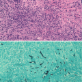
Fungal colonization is usually found in patients with impaired mucociliary transport, particularly on nasal crusts. A fungus ball is a noninvasive, dense conglomeration of fungus hyphae mostly found in the maxillary sinus without tissue invasion but sometimes accompanied by a weak noneosinophilic mucosal inflammatory response [7].
Bent and Kuhn in 1994 published the diagnostic criterion for fungal rhinosinusitis, which is largely regarded as the standard for diagnosis today. Patients must meet all the major criteria for diagnosis, while the minor criteria serve to support the diagnosis and describe individual patients but are not used to make a diagnosis. The major criteria include a history of type I hypersensitivity by history, skin testing, or in vitro testing, nasal polyposis, characteristic computed tomography (CT) scan findings, the presence of eosinophilic mucin without invasion, and a positive fungal stain of sinus contents removed at the time of surgery. The minor criteria include a history of asthma, unilateral predominance of disease, radiographic evidence of bone erosion, fungal cultures, presence of Charcot-Leyden crystals in surgical specimens, and serum eosinophilia [8].
Moreover, special forms of chronic rhinosinusitis exist often referred to as eosinophilic fungal rhinosinusitis (EFRS), including allergic fungal rhinosinusitis.
EFRS is a noninvasive chronic eosinophilic sinus inflammation frequently associated with nasal polyps. A characteristic of eosinophilic fungal rhinosinusitis is the presence of highly viscid sinus secretions with eosinophil decay products, termed eosinophilic mucus by Bent and Kuhn. EFRS may be further divided into allergic fungal rhinosinusitis (AFRS) with a positive diagnostic test for IgE-mediated allergy to the fungal elements detected within the sinus. It is considered an IgE-mediated mucosal hypersensitivity directed against fungal antigens deposited on the sinus mucosa. If type I allergy tests to molds are negative but eosinophilic mucus with fungal elements is found, the term non-allergic EFRS is used [9, 10].
Other Forms of Fungal Sinus Disease
Acute invasive fungal rhinosinusitis almost exclusively occurs in immunocompromised hosts and is characterized by hyphal invasion of surrounding tissues, vascular invasion, and tissue necrosis. Today, mortality ranges between 20 and 50 % and is mainly dependent on the improvement of the immunity of the host [11, 12]. Chronic invasive fungal rhinosinusitis is a non-granulomatous, slowly destructive process with abundant hyphae on histopathologic examination. Chronic invasive fungal rhinosinusitis is commonly seen in patients with less severe immune dysfunction like diabetes mellitus and corticosteroid treatment. A 40 % mortality rate has been reported. In South Asia a granulomatous invasive fungal rhinosinusitis with noncaseating granulomas around sparse hyphae is found. This form also has a significant mortality [11, 12].
Treatment of Fungus in CRS
Potential Indications for Oral and Topical Antifungals
In the rest of this chapter, we will mainly talk about treatment of fungus in patients with a normal immunity. At the end of the chapter, in a short paragraph we will talk about treatment of fungal sinus disease in immunocompromised patients.
As aforementioned, the main forms of fungal disease in patients with normal immunity are the fungus ball [7] and eosinophilic fungal rhinosinusitis [9].
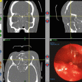
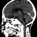
The fungus ball usually appears as a mass within the lumen and is usually unilateral and limited to one paranasal sinus. The maxillary sinus is most frequently affected, followed by the sphenoid sinus. The most common symptoms are purulent nasal discharge, facial pain or fullness, chronic nasal obstruction, fetid smell perception, and postnasal discharge. However, it is not uncommon for these lesions to be recognized as an incidental radiological finding in an asymptomatic patient. Fungus balls typically appear hyperdense on CT scans and frequently show calcifications [7, 13]. Sinus walls may be hypersclerotic or expanded and thinned. On T1-weighted MRI, a fungus ball appears hypointense. Calcifications and paramagnetic metals, such as iron, magnesium, and manganese, generate areas of signal void in T2-weighted images. The treatment of fungus balls is surgical. The goal of the surgery is to remove all fungal material without unnecessary damaging the mucosa. Additional medical treatment is not necessary. The recovery is usually excellent [14, 15].
Eosinophilic Fungal Rhinosinusitis
The concept of AFRS (a subtype of CRS) parallels allergic bronchopulmonary aspergillosis (ABPA), in which hypersensitivity reactions to Aspergillus species colonizing the lower respiratory tract result in significant pathology [1]. As clinical evidence for AFRS accumulated, controversy regarding its definition (should fungal allergy be present?), prevalence, and disease mechanisms emerged.
From the immunological point of view, patients with AFRS present (a) type I hypersensitivity to multiple molds and non-fungal aeroallergens demonstrable by immediate skin test reactions and in vitro detection of sIgE, although sensitization rates to fungi do not seem to be higher in patients with AFRS than in patients with other forms of CRS [2] or patients with allergic rhinitis [16]; (b) increased total IgE, which can also be found in the absence of sensitization to fungus at all or sensitization to other fungi than the ones found in the sinus [17]; and (c) increased sIgG to multiple molds [16]. Fungal-specific precipitins and peripheral eosinophilia are presented inconsistently.
Although the exact relevant pathophysiological mechanisms are unclear and widely discussed, we can summarize that it should be questioned whether a type I hypersensitivity to fungi is relevant for the development of CRS, even AFRS. Whether fungal-specific IgG (especially IgG1 and IgG3) is involved in the pathogenesis of CRS requires additional research.
Fungus Anti-host Effects
Besides innate and adaptive antifungal immune responses that may contribute to disease development, fungus anti-host effects may be involved in CRS pathogenesis. Ubiquitous airborne fungi (especially Alternaria and Aspergillus) are known to produce proteases that bind to protease-activated receptors (PARs) expressed on epithelial cells, airway cells, leukocytes, and blood vessels, thereby activating intracellular signaling pathways that give rise to multiple responses, including the production and release of mediators involved in tissue damage [18, 19].
In addition to an indirect effect, Alternaria alternata may activate eosinophils directly. Alternaria alternata, but not IL-5, has been shown to induce eosinophil IL-8 synthesis and eosinophil surface expression of CD11b (a β2-integrin that is used by eosinophils to adhere to β-glucan, a major fungal cell wall component [20]) and CD63 (a component of eosinophil granule membranes) in healthy volunteers, patients with allergic rhinitis, and patients with bronchial asthma.
Upon recognition of Alternaria alternata, eosinophil-released eosinophil-derived neurotoxin (EDN) [21] may play a pivotal role in CRS pathogenesis.
Specific Therapy
Oral Antifungals
Related posts:

Stay updated, free articles. Join our Telegram channel

Full access? Get Clinical Tree


