Supratarsal crease fixation in the Asian patient can provide a more open-eyed, awake look without compromising their ethnic appearance. A conservative supratarsal crease height and conservative to no removal of postseptal fat help to ensure this natural-appearing result. With the full-incision method, consistently excellent results have been achieved with durable crease fixation despite a prolonged recovery time. The supratarsal crease fixation provides an excellent method for the younger patient seeking cosmetic eyelid enhancement. However, for the aging Asian patient, the complexity of the strategy is greater.
What makes blepharoplasty on Asian patients unique is the management of the supratarsal crease. Although the presence of a supratarsal crease is a naturally occurring anatomic finding in the Asian population, many of those who lack this anatomic trait will often seek surgical creation de novo . The desire to have a double eyelid is largely cultural, as this feature is considered attractive.
The primary goal of this procedure is not only to create a supratarsal crease but also to create a crease that is consistent with the natural configuration present in the population. From a surgical point of view, this requires a thorough understanding of the natural crease shape and characteristic and mastery of the unique skills required to create it. Asian upper-eyelid blepharoplasty has a rich and complex history. The first reported case was performed and reported in the late nineteenth century. Since then, several innovative surgeons began to describe their techniques and concepts. The era of westernization upper blepharoplasty, which focused on creating a high supratarsal crease consistent with the White norm, has given way to methods that preserve ethnic characteristics. The current strategies can be broadly categorized into suture-based, full-incision, and partial-incision techniques.
The method that is advocated in this article is the full-incision technique. The rationale for this preference can be summarized as follows:
- (1)
Relative permanence compared with other methods
- (2)
No need to rely on any buried permanent sutures to hold the fixation
- (3)
Ease in identifying postseptal tissues through a wider aperture
- (4)
Ability to modulate excessive skin (dermatochalasis) in the aging eyelid.
The major drawback of the full-incision method is the protracted recovery time, in which the patient can look grossly abnormal for 1 to 2 weeks, and still not entirely natural for months, if not a full year. Scarring has proven to be a nonissue if the delicate tissues near the epicanthus are carefully avoided. In the authors’ opinion, the incision line is more difficult to observe with the full-incision than with the partial-incision method because there is no abrupt ending as is apparent with the more limited-incision technique.
Operative technique
Determination of the Eyelid Crease Position
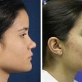
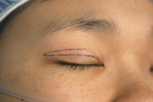
The first step is designing the proposed eyelid crease. There are several variations ranging from inside fold (the medial incision terminates lateral to the epicanthus) and outside fold (the medial incision extends medial to the epicanthus by 1–2 mm). There are 2 variations to the shape of the incision. The first is an oval shape (slight flare of the crease height laterally above the ciliary margin) versus rounded, in which the line runs parallel to the ciliary margin. Our preference is for the inside fold paired with an oval configuration ( Fig. 1 ).
Surgical Marking
The patient should be placed in the supine position and the upper-eyelid skin is held taut to the point that the eyelashes are just beginning to evert. The supratarsal crease should be marked at a distance of 7 mm from the ciliary crease to create a natural, low crease design (which constitutes the naturally occurring shape). The degree of skin excision to be performed should err on the side of conservatism with about 3 mm between the upper and lower limbs.
Anesthesia
Deep sedation should be avoided, as patient cooperation is vital to ensure symmetry toward the end of the procedure. A mixture of 0.5 mL of 1% lidocaine with 1:100,000 epinephrine and 0.5 mL of 0.25% bupivicaine with 1:100,000 epinephrine attached to a 30-gauge needle is used to infiltrate the upper-eyelid skin by raising 2 to 3 subcutaneous wheals, which are then manually distributed by pinching the skin along the entire length of the incision. This method avoids threading the needle and limits the chance of a hematoma that can lead to difficulty in gauging symmetry during the procedure. A total of only 1 mL of the local anesthesia mixture described earlier is infiltrated along each proposed incision to maintain symmetry.
Surgical Exposure
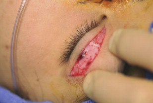
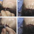
A no.15 blade is used to incise the skin down through the orbicularis oculi muscle, taking care not to pass the blade much further than that initial depth ( Fig. 2 ). Bipolar cautery is used to coagulate the vascular arcades that run perpendicularly across the incision line to limit unnecessary bleeding and thereby mitigate swelling and distortion during this delicate procedure. The depth of the incision can be further deepened with the no.15 blade down toward the orbital septum before removing the skin island with curved iris scissors. Additional cautery is used as needed. At this point, the same procedure is performed on the contralateral side and is continued in this alternating fashion to ensure symmetry.