Stiffness
Hill Hastings II
Jeffry B. Watson
Surasak Jitprapaikulsarn
Digital Stiffness
Congruent articular surfaces, tendon mobility, muscular strength, and a pliable soft tissue envelope are prerequisites to digital motion. Digital stiffness results from processes that compromise one or more of these factors. The etiology of digital stiffness is wide ranging and includes Dupuytren contracture, central or peripheral nervous system disorders, congenital disorders such as camptodactyly or arthrogryposis, and posttraumatic or rheumatologic alteration in regional bone morphology or joint stability.
This chapter will focus on stiffness resulting from trauma, swelling, or immobilization without change in periarticular bone morphology.
I. Pathoanatomy
Hand stiffness commonly results from a combination of prolonged edema, ischemic muscle contracture, and direct tendon scarring.
Contractures typically become fixed with the MP joints in extension, the interphalangeal joints in flexion, and the thumb metacarpal in adduction.
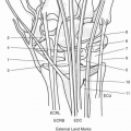
MP joint
The metacarpal head is trapezoidal with a wider palmar than dorsal surface.
As the MP collateral ligaments originate in the retrocondylar recess, passing volarly to insert on the palmar aspect of the proximal phalanx, the collateral ligaments are taut in flexion. This “cam” effect results in greater stability of the MP joint in flexion (coupled with a larger joint surface contact area).
Provided greater joint laxity in extension, the patient with an edematous digit will most comfortably rest in MP extension. Over time, the collateral ligaments become fixed and are unable to lengthen as necessary to permit MP flexion.
PIP joint
Unlike the MP joint, the PIP joint has no “cam” effect and the fluid capacity of the joint is similar in flexion and extension. Also, the PIP volar plate is thicker, less pliable, and does not “telescope” in length like the MP volar plate.
The tendency for the PIP joint to become stiff in flexion is explained by the relative increased tension in the extrinsic flexors due to the MP joint extension. Chronically, the volar plate and contracted collaterals can contribute to the inability to extend the PIP joint.
In addition to losing terminal extension, PIP joints also tend to lose terminal flexion. Extremes of flexion are sacrificed with persistent dorsal soft tissue edema. Normally, the dorsal PIP joint skin requires 12 mm of lengthening to obtain 90 degrees of flexion. With 5 mm of edema over the joint, the
same degree of flexion requires 19 mm of lengthening in skin that is now less pliable. Over time, skin, collateral ligament, and dorsal capsule contractures combine to create fixed contractures that limit PIP flexion as well.
DIP joint
DIP joint structure is similar to that of the PIP joint, with broad articular contact area, a central articular groove, and equal collateral ligament tension throughout the arc of motion.
The volar plate is different from that of the PIP joint due to the absence of checkrein ligaments to prevent hyperextension.
Extensor mechanism
The digital extensor mechanism is divided into the intrinsic and extrinsic components.
Extrinsic extensors (EIP, EDQ, and EDC) originate proximal to the wrist. These tendons cross the dorsal midline of the MP joint to become the central extensor slip inserting at the PIP joint. Note that the extrinsic extensors are the sole mechanism for MP extension by means of their action through their attachment to the volar plate through the sagittal bands. Short, direct fiber attachments from the undersurface of the tendon to the dorsal capsule and dorsal phalangeal base are described, but these do not have constant effect on MP extension. Further distal, the long extrinsic extensor sends off two lateral slips to merge with the lateral bands of the intrinsic system in the distal portion of the proximal phalanx to form the conjoined lateral band.
Intrinsic extensor mechanism
The intrinsic extensors comprise seven interosseous (four dorsal and three volar) muscles and four lumbricals. The deep motor branch of the ulnar nerve innervates all interossei and the lumbricals to the ring and small fingers. The long and index lumbricals are median nerve innervated (via fibers emanating from the common digital nerves to the first and second webspaces).
Interossei—Flex the MP and extend the IP joints (the “intrinsic plus” posture).
Dorsal interossei abduct the digits from the middle finger.
Volar interossei adduct the digits toward the middle finger.
The superficial head of the dorsal interossei inserts on the proximal phalanx providing abduction and secondarily MP flexion. The deep head of the dorsal interossei pass dorsal to the deep transverse metacarpal ligament (DTML) (but volar to the axis of rotation of the MCP joints) to insert into the lateral bands, which immediately send transverse fibers dorsally toward the central extensor tendon to contribute to MP flexion. Just distally, the oblique fibers arch distally and dorsal to insert alongside the central slip on the dorsal lateral tubercles of the middle phalangeal base to effect PIP extension.
The lateral bands are joined by the lumbricals (which have just passed palmar to the DTML) on the radial aspect of each digit and lateral slips of the central extensor tendon to form the conjoined lateral band. The conjoined lateral bands from each side of the digit merge over the middle phalanx to form the terminal tendon, which extends the DIP joint.
The volar interossei have no direct attachment to the proximal phalanx and form the lateral band on the opposite side of the
digit from the lateral band formed by the dorsal interossei, with identical contributions of transverse and oblique fibers.
Contracture of the interosseous muscles limits simultaneous MP extension and IP flexion. This “intrinsic tightness” is a frequent occurrence following prolonged edema in the hand or in situations following vascular compromise, such as a compartment syndrome.
The lumbricals
The four lumbricals originate on the FDP tendons in Zone 3 of the palm, with the index and long (median innervated) arising only from the FDP to those fingers and the ring and small (ulnar innervated) arising from the FDP tendons from the adjacent digits as well. Unlike the interosseous tendons, the lumbrical tendons pass palmar to the DTML before joining the lateral band to become the conjoined tendon. Lumbrical contraction relaxes the FDP tendon (its origin and antagonist) while creating tension in the lateral band to extend the IP joints. The volar position of the tendon relative to the DTML at the MP joint further contributes to MP flexion.
Any abnormal tension of the lumbricals will affect IP joint motion. As with the interossei, this can result from muscle ischemia, decreasing the excursion of the lumbrical muscle-tendon unit. Direct scarring of the lateral band tendon to the bone (often seen following phalangeal fracture) will also limit lateral band excursion and IP motion. The tendon of the lumbrical has also been known to scar down to the interosseous tendon, forming an adhesion that “straddles” the DTML and limits excursion of both.
Flexor tendons
The retraction of avulsed or unrepaired FDP tendon can also result in limited flexion of the PIP joint, despite the FDS tendon being intact. As the FDP retracts, the lumbrical origin is moved proximally, increasing the lumbrical’s resting tension. Attempted flexion results in further FDP retraction and tension through the “pretensioned” lumbrical tendon, placing a paradoxial extension moment at the PIP (because of the lateral bands situated dorsal to the axis of rotation of the PI joint) and resisting flexion from the FDS. This is known as the lumbrical plus deformity.
Skin
The skin of the hand and digit contributes to stiffness in several ways. Acutely, edematous skin necessitates an increased force to accomplish motion. Chronically, skin becomes contracted around stiff joints contributing to contracture. Traumatic and surgical wounds can also produce joint contractures or scar to flexor and extensor mechanisms to limit motion.
II. Evaluation of the Stiff Digit
This discussion assumes that all skeletal issues such as phalangeal nonunion or malunion, intraarticular incongruity, and periarticular bony blocks to motion have been corrected.
Barriers to flexion include dorsal capsular contracture, contracted collateral ligaments, adherent extensor tendons, dorsal skin contracture (such as wounds allowed to heal by secondary intention), and intrinsic tightness.
Flexor tendon adhesions, rupture or excessive repair site gap will impair active flexion while still allowing passive flexion.
Extension blocks include volar capsule (volar plate) contracture, palmar skin contractures, and adherent flexor tendons.
Insufficiency or scarring of extrinsic extensor motor units will impair active while still permitting passive extension.
Identification of all components limiting active joint motion may occur only after fixed joint contractures are resolved, allowing assessment of the integrity of the flexor and extensor motor tendon units.
Evaluation begins with comparison of active motion versus passive motion of both the PIP and MP joints.
If passive motion is greater, then the most limiting problem is outside of the joint itself. Specifically, the flexor or extensor motor unit is either denervated, incompetent or tethered by scar.
Similarly, if passive motion changes with the position of surrounding joints, the etiology is outside of the joint itself.
When the intrinsics have a normal degree of elasticity and excursion without other factors blocking MP or PIP motion, one can easily assume the intrinsic minus position of full MP extension and IP flexion.
When there is diminished excursion of the intrinsics, they cannot stretch enough to allow for the full intrinsic minus position. That is, PIP flexion can only be achieved through compromise of MP extension, and MP extension can only occur if the PIP is extended. This is referred to as intrinsic tightness. Tightness of the lumbricals relative to the interossei can further be differentiated by passively flexing the PIP joint and DIP joint together. If resistance is noted with passive DIP flexion despite facile PIP flexion during the Bunnell intrinsic tightness test, the lumbrical muscle may be the contracted component.
Extrinsic tightness occurs when there is diminished passive excursion of the EDC, EIP, or EDQ muscle-tendon units. The most common cause of this is when the tendon is adherent to surrounding structures following trauma. In this situation, simultaneous flexion of the MP and PIP joints is limited. The differentiation of extrinsic versus intrinsic tightness blocking flexion is key to developing any treatment plan.
Oblique retinacular ligament contracture is detected by passive simultaneous extension of the DIP and flexion of the PIP joints (Boutonniere). This structure acts as a passive link for simultaneous flexion and extension of the PIP and DIP joints. If the ligament is contracted, DIP flexion will only occur with some degree of PIP flexion to relax the ligament.
III. Treatment
Nonoperative therapeutic options should be exhausted prior to consideration for surgical treatment.
The results of a sustained progression through splinting and motion are generally enduring with less potential morbidity.
Splinting regimens that stretch contracted joints or tissues operate on the principle that sustained tension changes tissues elasticity and cell proliferation.
Dynamic splints apply stretch through springs or elastic bands against contracted tissues while still allowing for some motion.
Static splints maintain the joint in one position without applying force, and serial static splints hold the joint in one position while applying stretch to contracted tissues. As the contracted tissues relax in response, the serial static splint is remolded or changed to continually apply stretch against the contracture while being worn. Static progressive splints operate on the same principle, but they have some component (such as a gear or ratchet) that allows the splint position to be modified without custom molding.
Treatment goals should be individualized for the digit and the patient. Some patients may be able to function without full digital flexion of both hands and are less willing to undergo extreme or prolonged measures to obtain that. Furthermore, the ulnar-sided digits generally require a greater arc of motion than the long and index fingers, whose tasks are mostly performed in conjunction with the thumb and require only moderate degrees of flexion.
As noted above, MP joints should be immobilized in flexion. However, if stiff in extension, a program of dynamic and static progressive splinting combined with aggressive hand therapy usually results in significant improvement of motion.
Depending on the initiating pathology, the PIP joint can exhibit limited flexion, extension, or both. Regimens of daytime dynamic splinting with nighttime serial static splinting and stretching exercises frequently result in improvement.
Surgical release can be considered when nonoperative results have plateaued and are not adequate for function. Furthermore, several key criteria must be satisfied.
All soft tissue swelling and inflammation should be resolved.
Wounds should be healed or covered beforehand.
Articular surfaces should be stable and congruent. Release of a noncongruent joint usually results in temporary instability followed by stiffness.
Finally, the patient needs to be fully informed and committed to a prearranged aggressive postoperative motion therapy regimen that begins from postoperative day two or three. Only if these conditions are met should surgical release be scheduled.
After determining the amount of active and passive motion in each direction at each joint, Boyer and colleagues have outlined a systematic operative approach for six combinations of stiffness based on physical examination findings.
No passive flexion or extension. This suggests both dorsal and volar pathology.
Dorsal joint contracture, contracture of dorsal portion of collate ral ligaments, or extensor adhesions can inhibit gliding of these structures and limit joint flexion.
On the palmar aspect, accessory collateral ligament contracture, volar plate checkrein ligament contracture, skin contracture, and flexor tendon adhesions will limit passive extension.
Dorsal and volar procedures performed during the same anesthetic are not advisable, as flap necrosis or prohibitive pain and swelling often result and undermine intraoperative gains. Therefore, the volar aspect is usually addressed first in a progressive sequence as below.
Two or four flap Z-plasties in skin
Checkrein ligament, accessory collateral release
Palmar or complete collateral release
Provision of stable gliding skin surface
Following restoration of full passive extension and subsidence of postoperative swelling and inflammation, dorsal release can be pursued to restore passive flexion and also the active component of extension.
Recalling the preoperative assessment to check for intrinsic versus extrinsic extensor tightness.
If intrinsic tightness is present, an intrinsic release will need to be performed by resecting a wedge of tendon from the lateral bands on each side of the digit.
In the setting of extrinsic tightness or dorsal joint contracture, sequential dorsal release is needed. Working on either side of the central slip tendon (it does not need to be disrupted), the dorsal joint capsule is resected, and any adhesions between the bone and the extensor tendon are excised. Next, the dorsal portion of the collateral ligament origins should be resected, followed by the remainder of the origins if passive flexion is still incomplete.Related posts:

Stay updated, free articles. Join our Telegram channel

Full access? Get Clinical Tree