Summary
Split-thickness skin grafting can provide large volumes of color-matched skin for head and neck reconstruction with minimal donor-site morbidity.
Split-thickness skin graft harvesting from the scalp provides thick color-matched skin with the advantage of the donor site remaining in the operative field.
There is little role for meshing split-thickness skin grafts in head and neck reconstruction given the final aesthetic result is unacceptable.
6.1 General Considerations
The usefulness of split-thickness skin grafting (STSG) in head and neck reconstruction may well be more than generally appreciated and is greatly expanded by STSG in conjunction with dermal regeneration templates. 1 The ease and reliability of split-thickness grafting and the ability to provide large volumes of thin color-matched skin that will reliably heal to an excellent cosmetic result with very acceptable donor-site appearance makes it a very valuable technique for head and neck reconstruction. 2 The decision to use either a split-thickness color-matched skin graft or a full-thickness color-matched skin graft for head and neck reconstruction depends on the quality of the skin, the size requirements of the skin, as well as considerations regarding both harvest and donor-site management.
Historically, many surgeons feel that general anesthetic is required for safe harvest of STSG; however, in our practice at the UTWSMC, we have comfortably and easily performed STSG under both local and intravenous sedation. Limiting the use of anesthesia is safer for elderly patients who may not be suitable candidates for general anesthesia.
6.2 Donor-Site Selection
The selection of donor site begins with the determination of whether color-matched skin is required and following the same principles as for full-thickness skin grafting, if color-matched skin is required then the graft must be harvested above the clavicles. In many ways, the scalp is an ideal donor site for STSG. 3, 4 It is very thick, very vascular, and provides a robust graft and if harvested appropriately on shaved hair-bearing scalp, the donor site heals very rapidly and can heal with a nearly imperceptible scar. 3, 4, 5 Contrast this versus the very notable scar that is left following harvest of STSG from the traditional site on the leg.
For defects that do not require color-matched skin, the lateral or proximal thigh donor site is very accessible, very easy to harvest from, and is able to provide large volumes of smooth, non–color-matched skin; however, it can leave an unsightly scar, particularly on young females, that is difficult to cover. 6 It is preferable to have the patient wear an appropriate underwear or swimsuit bottom and then mark out the borders of the clothing and then take the graft within the borders. For current fashions, this usually results in a graft that is harvested medially from a single buttock. Although this will result in painful donor-site sitting for 5 to 10 days, it does result ultimately in a scar that can be completely hidden by clothing. 6
6.3 Harvest Technique
After selection of the skin graft harvest site, careful measurement of the required graft dimensions and careful infiltration of the donor site are performed with local anesthetic to provide hemostasis, pain control, and some degree of turgor to the skin that will aid in the graft harvest. The selection of the Zimmer dermatome blade guard is also important, given that oftentimes the 3-in. guard is the only template that will harvest the entire dimensions of the guard. A 4-in. guard, although it will harvest larger than 3 in., will result in the harvested skin not fitting on the standard meshing carriers. This is also true of the 2-in. guard that will also oftentimes harvest less than the 2-in. guard dimensions. The decision on harvested skin graft thickness involves three factors: (1) the thicker the harvested graft, the longer the donor-site healing—and with elderly thin skinned patients, it is possible to inadvertently harvest skin thick enough to result in a full-thickness donor-site defect; (2) the thicker the graft, the more prolonged the graft healing; and (3) thicker grafts heal with less primary and secondary skin contracture. Additionally, even though the manufacturer recommends for Integra graft of 0.008 thickness, our clinical experience with Integra is that a robust Integra base is able to heal much thicker skin grafts, even up to 0.012 thick, which is more easily harvested and easier to handle during inset. 7
Several considerations are to be made in setting up a dermatome for graft harvest. This includes the selection of the blade guard and ensuring the blade has not been inadvertently placed upside down. Even though there are safeguards in place on the dermatome itself to prevent incorrect blade placement, it is still a possible mistake and will result in the blade guard being ineffective, resulting in an instantaneous full-thickness skin defect when graft harvest is attempted. Care must be taken that a Padgett blade is also not placed into a Zimmer dermatome, because this will result in no protection by the guard and a full-thickness injury to the donor site. In setting up the donor site after infiltration of local anesthetic, care must be taken that the drapes do not interfere with the full path of the harvest and is useful to perform a practice pass without power on the dermatome to ensure there are no obstructions to harvesting. A very common practice of using a no. 15 blade as a “feeler gauge” to measure the blade thickness is infeasible, is not accurate, and is not recommended by the Zimmer dermatome manuals. This is a habit that is easily discarded with no ill effect for the patient. Mineral oil is routinely used on the guard, as well as on the donor site. After the graft is harvested, initial attention is to be focused on the donor site. There is literature in support of an inclusive dressing of either Tegaderm or BioDerm to be placed directly over the donor site and great care is taken for the accurate placement of this dressing with wide borders and use of a tissue adhesive on the normal skin borders to maintain this dressing in place. 8 The patient is given postoperative instructions to leave the adhesive in place for 5 to 7 days and healing can occur underneath. If the patient develops a large area of ballotable fluid unexpectedly, we encourage them to make a small piercing on the Tegaderm with nail scissors and drain it and make every attempt to leave the occlusive Tegaderm dressing in place until the donor site heals underneath. For surgeons who are interested in the final aesthetic outcome, some degree of insight needs to go into the decision-making process of actually meshing the graft. Although meshing does increase the volume of usable skin, and also likely increases the graft take, it will result in an irretrievably abnormal aesthetic result that is impossible to correct. The common pebble-stoning or screen door appearance of a meshed skin graft is unmistakable and is caused by the healing of the meshed interstices by scarring and not actual primary healing and results with a far inferior result than a unmeshed sheet graft. Additionally, the practice of meshing, but not expanding, also results in wound healing that is inferior to an appropriately inset unmeshed sheet graft.
6.4 Graft Inset
The inset process for large sheet graft relies on continuous fast or plain gut sutures (never chromic gut) along the border of the graft and then dependent on the size, as well as the eventual contours, small areas of the graft, where it is not adhered or where there are clear air bubbles underneath which are then sharply incised with a no. 69 Beaver blade and a 5–0 gut suture is utilized to secure one limb of the interstices to keep this open and allow the easy egress of any potential seroma fluid. The graft is then carefully bolstered with a surgical sponge bolster with ointment on one side and no intervening nonstick gauze. For large areas of graft, central 3–0 double-armed Prolene sutures are utilized to improve uniform compression of the sponge and the remainder of the sponge is sewn in place with 4–0 pop-off Nurolon sutures with no attempts to do a tie-over bolster, rather utilizing just peripherally placed sutures. The advantage of peripherally placed suture is the speed of the bolster inset and also that it prevents the peripheral upturning that can occur with tie-over bolsters. Postoperatively, the sponge is kept dry and removed in 5 to 7 days. On bolster removal and if there is any accumulation of seroma fluid, then this is carefully expressed on the initial phase and the graft recompressed and given a chance to continue healing (▶ Fig. 6.1, ▶ Fig. 6.2, ▶ Fig. 6.3, ▶ Fig. 6.4, ▶ Fig. 6.5, ▶ Fig. 6.6, ▶ Fig. 6.7, ▶ Fig. 6.8, ▶ Fig. 6.9, ▶ Fig. 6.10, ▶ Fig. 6.11, ▶ Fig. 6.12, ▶ Fig. 6.13, ▶ Fig. 6.14, ▶ Fig. 6.15, ▶ Fig. 6.16, ▶ Fig. 6.17, ▶ Fig. 6.18, ▶ Fig. 6.19, ▶ Fig. 6.20).
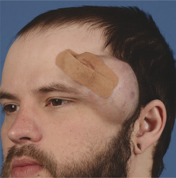
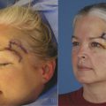
Fig. 6.1 A 32-year-old male patient with very large dermatofibrosarcoma protuberans tumor of left scalp.
Related posts:
Stay updated, free articles. Join our Telegram channel

Full access? Get Clinical Tree


